5DQF

Horse Serum Albumin (ESA) in complex with Cetirizine


Experimental Data Snapshot

  • Method: X-RAY DIFFRACTION
  • Resolution: 2.15 Å
  • R-Value Free: 0.241 
  • R-Value Work: 0.189 
  • R-Value Observed: 0.191 

wwPDB Validation   3D Report Full Report


Ligand Structure Quality Assessment 


This is version 1.8 of the entry. See complete history


Literature

Crystal structure of equine serum albumin in complex with cetirizine reveals a novel drug binding site.

Handing, K.B.Shabalin, I.G.Szlachta, K.Majorek, K.A.Minor, W.

(2016) Mol Immunol 71: 143-151

  • DOI: https://doi.org/10.1016/j.molimm.2016.02.003
  • Primary Citation of Related Structures:  
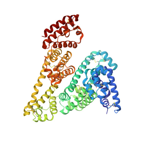
    5DQF

  • PubMed Abstract: 

    Serum albumin (SA) is the main transporter of drugs in mammalian blood plasma. Here, we report the first crystal structure of equine serum albumin (ESA) in complex with antihistamine drug cetirizine at a resolution of 2.1Å. Cetirizine is bound in two sites--a novel drug binding site (CBS1) and the fatty acid binding site 6 (CBS2). Both sites differ from those that have been proposed in multiple reports based on equilibrium dialysis and fluorescence studies for mammalian albumins as cetirizine binding sites. We show that the residues forming the binding pockets in ESA are highly conserved in human serum albumin (HSA), and suggest that binding of cetirizine to HSA will be similar. In support of that hypothesis, we show that the dissociation constants for cetirizine binding to CBS2 in ESA and HSA are identical using tryptophan fluorescence quenching. Presence of lysine and arginine residues that have been previously reported to undergo nonenzymatic glycosylation in CBS1 and CBS2 suggests that cetirizine transport in patients with diabetes could be altered. A review of all available SA structures from the PDB shows that in addition to the novel drug binding site we present here (CBS1), there are two pockets on SA capable of binding drugs that do not overlap with fatty acid binding sites and have not been discussed in published reviews.


  • Organizational Affiliation

    Department of Molecular Physiology and Biological Physics, University of Virginia, Charlottesville, VA 22908-0736, USA; New York Structural Genomics Research Consortium (NYSGRC), USA.


Macromolecules
Find similar proteins by:  (by identity cutoff)  |  3D Structure
Entity ID: 1
MoleculeChains Sequence LengthOrganismDetailsImage
Serum albumin580Equus caballusMutation(s): 1 
UniProt
Find proteins for P35747 (Equus caballus)
Explore P35747 
Go to UniProtKB:  P35747
Entity Groups  
Sequence Clusters30% Identity50% Identity70% Identity90% Identity95% Identity100% Identity
UniProt GroupP35747
Sequence Annotations
Expand
  • Reference Sequence
Small Molecules
Ligands 5 Unique
IDChains Name / Formula / InChI Key2D Diagram3D Interactions
CZE
Query on CZE

Download Ideal Coordinates CCD File 
N [auth A](2-{4-[(S)-(4-chlorophenyl)(phenyl)methyl]piperazin-1-yl}ethoxy)acetic acid
C21 H25 Cl N2 O3
ZKLPARSLTMPFCP-NRFANRHFSA-N
LCR
Query on LCR

Download Ideal Coordinates CCD File 
M [auth A](2-{4-[(R)-(4-chlorophenyl)(phenyl)methyl]piperazin-1-yl}ethoxy)acetic acid
C21 H25 Cl N2 O3
ZKLPARSLTMPFCP-OAQYLSRUSA-N
PG4
Query on PG4

Download Ideal Coordinates CCD File 
B [auth A]
C [auth A]
D [auth A]
E [auth A]
F [auth A]
B [auth A],
C [auth A],
D [auth A],
E [auth A],
F [auth A],
G [auth A],
H [auth A]
TETRAETHYLENE GLYCOL
C8 H18 O5
UWHCKJMYHZGTIT-UHFFFAOYSA-N
SO4
Query on SO4

Download Ideal Coordinates CCD File 
I [auth A],
J [auth A],
K [auth A],
L [auth A]
SULFATE ION
O4 S
QAOWNCQODCNURD-UHFFFAOYSA-L
CL
Query on CL

Download Ideal Coordinates CCD File 
O [auth A],
P [auth A],
Q [auth A],
R [auth A]
CHLORIDE ION
Cl
VEXZGXHMUGYJMC-UHFFFAOYSA-M
Binding Affinity Annotations 
IDSourceBinding Affinity
CZE Binding MOAD:  5DQF Kd: 2.53e+5 (nM) from 1 assay(s)
LCR Binding MOAD:  5DQF Kd: 2.53e+5 (nM) from 1 assay(s)
Experimental Data & Validation

Experimental Data

  • Method: X-RAY DIFFRACTION
  • Resolution: 2.15 Å
  • R-Value Free: 0.241 
  • R-Value Work: 0.189 
  • R-Value Observed: 0.191 
  • Space Group: P 61
  • Diffraction Data: https://doi.org/10.18430/M3WC7F
Unit Cell:
Length ( Å )Angle ( ˚ )
a = 94.234α = 90
b = 94.234β = 90
c = 141.893γ = 120
Software Package:
Software NamePurpose
MD2data collection
HKL-3000data reduction
DENZOdata reduction
SCALEPACKdata scaling
MOLREPphasing
Cootmodel building
REFMACrefinement
PDB_EXTRACTdata extraction

Structure Validation

View Full Validation Report


Ligand Structure Quality Assessment 


Entry History & Funding Information

Deposition Data


Funding OrganizationLocationGrant Number
National Institutes of Health/National Institute of General Medical Sciences (NIH/NIGMS)United StatesNIH-5U54GM094662-05

Revision History  (Full details and data files)

  • Version 1.0: 2015-12-23
    Type: Initial release
  • Version 1.1: 2016-02-24
    Changes: Other
  • Version 1.2: 2016-03-09
    Changes: Data collection, Database references
  • Version 1.3: 2017-09-27
    Changes: Author supporting evidence, Database references, Derived calculations, Refinement description
  • Version 1.4: 2018-08-15
    Changes: Data collection, Database references
  • Version 1.5: 2018-09-12
    Changes: Data collection, Database references
  • Version 1.6: 2019-12-25
    Changes: Author supporting evidence
  • Version 1.7: 2022-04-13
    Changes: Database references, Structure summary
  • Version 1.8: 2023-09-27
    Changes: Data collection, Refinement description