4H4F

Crystal structure of human chymotrypsin C (CTRC) bound to inhibitor eglin c from Hirudo medicinalis


Experimental Data Snapshot

  • Method: X-RAY DIFFRACTION
  • Resolution: 1.90 Å
  • R-Value Free: 0.210 
  • R-Value Work: 0.157 
  • R-Value Observed: 0.160 

wwPDB Validation   3D Report Full Report


This is version 1.2 of the entry. See complete history


Literature

Long-range Electrostatic Complementarity Governs Substrate Recognition by Human Chymotrypsin C, a Key Regulator of Digestive Enzyme Activation.

Batra, J.Szabo, A.Caulfield, T.R.Soares, A.S.Sahin-Toth, M.Radisky, E.S.

(2013) J Biol Chem 288: 9848-9859

  • DOI: https://doi.org/10.1074/jbc.M113.457382
  • Primary Citation of Related Structures:  
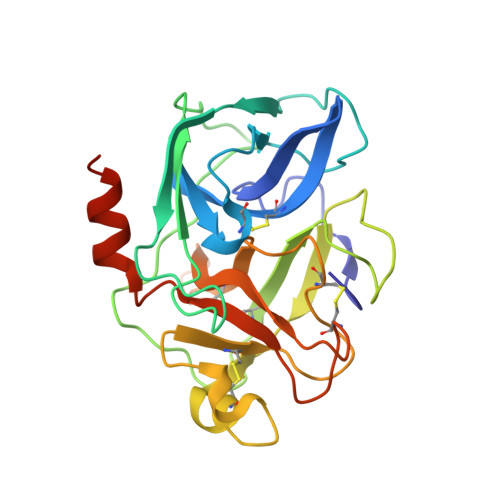
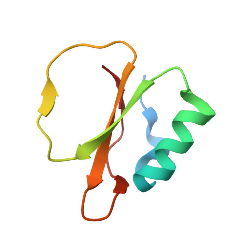
    4H4F

  • PubMed Abstract: 

    Human chymotrypsin C (CTRC) is a pancreatic serine protease that regulates activation and degradation of trypsinogens and procarboxypeptidases by targeting specific cleavage sites within their zymogen precursors. In cleaving these regulatory sites, which are characterized by multiple flanking acidic residues, CTRC shows substrate specificity that is distinct from that of other isoforms of chymotrypsin and elastase. Here, we report the first crystal structure of active CTRC, determined at 1.9-Å resolution, revealing the structural basis for binding specificity. The structure shows human CTRC bound to the small protein protease inhibitor eglin c, which binds in a substrate-like manner filling the S6-S5' subsites of the substrate binding cleft. Significant binding affinity derives from burial of preferred hydrophobic residues at the P1, P4, and P2' positions of CTRC, although acidic P2' residues can also be accommodated by formation of an interfacial salt bridge. Acidic residues may also be specifically accommodated in the P6 position. The most unique structural feature of CTRC is a ring of intense positive electrostatic surface potential surrounding the primarily hydrophobic substrate binding site. Our results indicate that long-range electrostatic attraction toward substrates of concentrated negative charge governs substrate discrimination, which explains CTRC selectivity in regulating active digestive enzyme levels.


  • Organizational Affiliation

    Department of Cancer Biology, Mayo Clinic Cancer Center, Jacksonville, Florida 32224.


Macromolecules
Find similar proteins by:  (by identity cutoff)  |  3D Structure
Entity ID: 1
MoleculeChains Sequence LengthOrganismDetailsImage
Chymotrypsin-C249Homo sapiensMutation(s): 0 
Gene Names: CLCRCTRC
EC: 3.4.21.2
UniProt & NIH Common Fund Data Resources
Find proteins for Q99895 (Homo sapiens)
Explore Q99895 
Go to UniProtKB:  Q99895
PHAROS:  Q99895
GTEx:  ENSG00000162438 
Entity Groups  
Sequence Clusters30% Identity50% Identity70% Identity90% Identity95% Identity100% Identity
UniProt GroupQ99895
Sequence Annotations
Expand
  • Reference Sequence
Find similar proteins by:  (by identity cutoff)  |  3D Structure
Entity ID: 2
MoleculeChains Sequence LengthOrganismDetailsImage
Eglin C70Hirudo medicinalisMutation(s): 0 
UniProt
Find proteins for P01051 (Hirudo medicinalis)
Explore P01051 
Go to UniProtKB:  P01051
Entity Groups  
Sequence Clusters30% Identity50% Identity70% Identity90% Identity95% Identity100% Identity
UniProt GroupP01051
Sequence Annotations
Expand
  • Reference Sequence

Find similar proteins by:  Sequence   |   3D Structure  

Entity ID: 3
MoleculeChains Sequence LengthOrganismDetailsImage
Chymotrypsin-CC [auth Q]10Homo sapiensMutation(s): 0 
Gene Names: CLCRCTRC
EC: 3.4.21.2
UniProt & NIH Common Fund Data Resources
Find proteins for Q99895 (Homo sapiens)
Explore Q99895 
Go to UniProtKB:  Q99895
PHAROS:  Q99895
GTEx:  ENSG00000162438 
Entity Groups  
Sequence Clusters30% Identity50% Identity70% Identity90% Identity95% Identity100% Identity
UniProt GroupQ99895
Sequence Annotations
Expand
  • Reference Sequence
Experimental Data & Validation

Experimental Data

  • Method: X-RAY DIFFRACTION
  • Resolution: 1.90 Å
  • R-Value Free: 0.210 
  • R-Value Work: 0.157 
  • R-Value Observed: 0.160 
  • Space Group: P 21 21 21
Unit Cell:
Length ( Å )Angle ( ˚ )
a = 56.272α = 90
b = 76.253β = 90
c = 81.823γ = 90
Software Package:
Software NamePurpose
SCALEPACKdata scaling
REFMACrefinement
PDB_EXTRACTdata extraction
HKL-2000data collection
DENZOdata reduction
PHASERphasing

Structure Validation

View Full Validation Report


Entry History 

Deposition Data

Revision History  (Full details and data files)

  • Version 1.0: 2013-02-27
    Type: Initial release
  • Version 1.1: 2013-03-06
    Changes: Database references
  • Version 1.2: 2013-04-24
    Changes: Database references