8QMO

Cryo-EM structure of the benzo[a]pyrene-bound Hsp90-XAP2-AHR complex


Experimental Data Snapshot

  • Method: ELECTRON MICROSCOPY
  • Resolution: 2.76 Å
  • Aggregation State: PARTICLE 
  • Reconstruction Method: SINGLE PARTICLE 

wwPDB Validation   3D Report Full Report


This is version 1.1 of the entry. See complete history


Literature

Structural Insights into the Activation of Human Aryl Hydrocarbon Receptor by the Environmental Contaminant Benzo[a]pyrene and Structurally Related Compounds.

Kwong, H.S.Paloni, M.Grandvuillemin, L.Sirounian, S.Ancelin, A.Lai-Kee-Him, J.Grimaldi, M.Carivenc, C.Lancey, C.Ragan, T.J.Hesketh, E.L.Balaguer, P.Barducci, A.Gruszczyk, J.Bourguet, W.

(2024) J Mol Biol 436: 168411-168411

  • DOI: https://doi.org/10.1016/j.jmb.2023.168411
  • Primary Citation of Related Structures:  
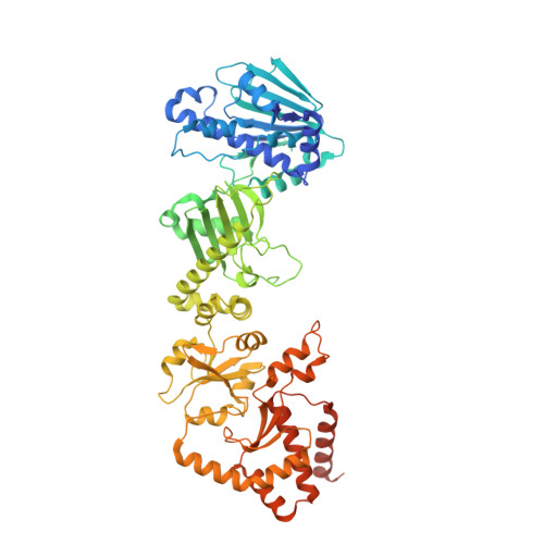
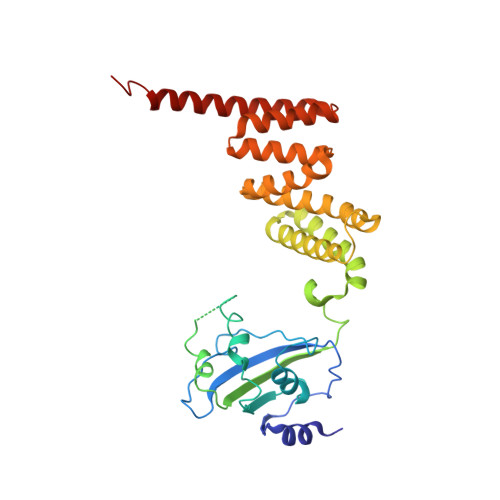
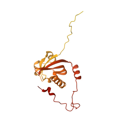
    8QMO

  • PubMed Abstract: 

    The aryl hydrocarbon receptor (AHR) is a ligand-dependent transcription factor belonging to the bHLH/PAS protein family and responding to hundreds of natural and chemical substances. It is primarily involved in the defense against chemical insults and bacterial infections or in the adaptive immune response, but also in the development of pathological conditions ranging from inflammatory to neoplastic disorders. Despite its prominent roles in many (patho)physiological processes, the lack of high-resolution structural data has precluded for thirty years an in-depth understanding of the structural mechanisms underlying ligand-binding specificity, promiscuity and activation of AHR. We recently reported a cryogenic electron microscopy (cryo-EM) structure of human AHR bound to the natural ligand indirubin, the chaperone Hsp90 and the co-chaperone XAP2 that provided the first experimental visualization of its ligand-binding PAS-B domain. Here, we report a 2.75 Å resolution structure of the AHR complex bound to the environmental pollutant benzo[a]pyrene (B[a]P). The structure substantiates the existence of a bipartite PAS-B ligand-binding pocket with a geometrically constrained primary binding site controlling ligand binding specificity and affinity, and a secondary binding site contributing to the binding promiscuity of AHR. We also report a docking study of B[a]P congeners that validates the B[a]P-bound PAS-B structure as a suitable model for accurate computational ligand binding assessment. Finally, comparison of our agonist-bound complex with the recently reported structures of mouse and fruit fly AHR PAS-B in different activation states suggests a ligand-induced loop conformational change potentially involved in the regulation of AHR function.


  • Organizational Affiliation

    CBS (Centre de Biologie Structurale), Univ Montpellier, CNRS, Inserm, Montpellier, France.


Macromolecules
Find similar proteins by:  (by identity cutoff)  |  3D Structure
Entity ID: 1
MoleculeChains Sequence LengthOrganismDetailsImage
Heat shock protein HSP 90-beta
A, B
730Homo sapiensMutation(s): 0 
Gene Names: HSP90AB1HSP90BHSPC2HSPCB
UniProt & NIH Common Fund Data Resources
Find proteins for P08238 (Homo sapiens)
Explore P08238 
Go to UniProtKB:  P08238
PHAROS:  P08238
GTEx:  ENSG00000096384 
Entity Groups  
Sequence Clusters30% Identity50% Identity70% Identity90% Identity95% Identity100% Identity
UniProt GroupP08238
Sequence Annotations
Expand
  • Reference Sequence
Find similar proteins by:  (by identity cutoff)  |  3D Structure
Entity ID: 2
MoleculeChains Sequence LengthOrganismDetailsImage
AH receptor-interacting protein330Homo sapiensMutation(s): 0 
Gene Names: AIPXAP2
UniProt & NIH Common Fund Data Resources
Find proteins for O00170 (Homo sapiens)
Explore O00170 
Go to UniProtKB:  O00170
PHAROS:  O00170
GTEx:  ENSG00000110711 
Entity Groups  
Sequence Clusters30% Identity50% Identity70% Identity90% Identity95% Identity100% Identity
UniProt GroupO00170
Sequence Annotations
Expand
  • Reference Sequence
Find similar proteins by:  (by identity cutoff)  |  3D Structure
Entity ID: 3
MoleculeChains Sequence LengthOrganismDetailsImage
Aryl hydrocarbon receptor439Homo sapiensMutation(s): 0 
Gene Names: AHR
UniProt & NIH Common Fund Data Resources
Find proteins for P35869 (Homo sapiens)
Explore P35869 
Go to UniProtKB:  P35869
PHAROS:  P35869
GTEx:  ENSG00000106546 
Entity Groups  
Sequence Clusters30% Identity50% Identity70% Identity90% Identity95% Identity100% Identity
UniProt GroupP35869
Sequence Annotations
Expand
  • Reference Sequence
Small Molecules
Ligands 4 Unique
IDChains Name / Formula / InChI Key2D Diagram3D Interactions
ADP
Query on ADP

Download Ideal Coordinates CCD File 
F [auth A],
I [auth B]
ADENOSINE-5'-DIPHOSPHATE
C10 H15 N5 O10 P2
XTWYTFMLZFPYCI-KQYNXXCUSA-N
W62 (Subject of Investigation/LOI)
Query on W62

Download Ideal Coordinates CCD File 
K [auth D]benzo[a]pyrene
C20 H12
FMMWHPNWAFZXNH-UHFFFAOYSA-N
MOO
Query on MOO

Download Ideal Coordinates CCD File 
E [auth A],
H [auth B]
MOLYBDATE ION
Mo O4
MEFBJEMVZONFCJ-UHFFFAOYSA-N
MG
Query on MG

Download Ideal Coordinates CCD File 
G [auth A],
J [auth B]
MAGNESIUM ION
Mg
JLVVSXFLKOJNIY-UHFFFAOYSA-N
Experimental Data & Validation

Experimental Data

  • Method: ELECTRON MICROSCOPY
  • Resolution: 2.76 Å
  • Aggregation State: PARTICLE 
  • Reconstruction Method: SINGLE PARTICLE 
EM Software:
TaskSoftware PackageVersion
RECONSTRUCTIONRELION3.1.1
MODEL REFINEMENTPHENIX1.20.1_4487

Structure Validation

View Full Validation Report


Entry History & Funding Information

Deposition Data


Funding OrganizationLocationGrant Number
ATIP-AvenirFranceR20059SP
Montpellier University of Excellence (MUSE)FranceANR-16-IDEX-0006
Other governmentEXPOAR23-RS03-JakubGruszczy
French National Research AgencyFranceANR-10-INBS-04-01

Revision History  (Full details and data files)

  • Version 1.0: 2024-01-10
    Type: Initial release
  • Version 1.1: 2024-02-28
    Changes: Database references