6HM1

Structural and thermodynamic signatures of ligand binding to an enigmatic chitinase-D from Serratia proteamaculans


Experimental Data Snapshot

  • Method: X-RAY DIFFRACTION
  • Resolution: 1.54 Å
  • R-Value Free: 0.203 
  • R-Value Work: 0.169 
  • R-Value Observed: 0.171 

wwPDB Validation   3D Report Full Report


Ligand Structure Quality Assessment 


This is version 2.1 of the entry. See complete history


Literature

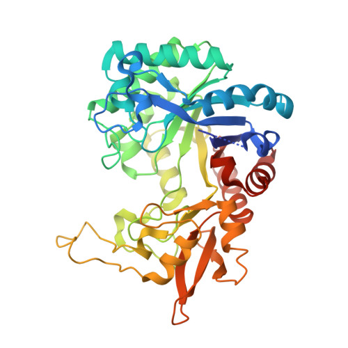
Structural and Thermodynamic Signatures of Ligand Binding to the Enigmatic Chitinase D of Serratia proteamaculans.

Madhuprakash, J.Dalhus, B.Vaaje-Kolstad, G.Sakuda, S.Podile, A.R.Eijsink, V.G.H.Sorlie, M.

(2019) J Phys Chem B 123: 2270-2279

  • DOI: https://doi.org/10.1021/acs.jpcb.8b11448
  • Primary Citation of Related Structures:  
    6HM1

  • PubMed Abstract: 

    The Gram-negative bacteria Serratia marcescens and Serratia proteamaculans have efficient chitinolytic machineries that degrade chitin into N-acetylglucosamine (GlcNAc), which is used as a carbon and energy source. The enzymatic degradation of chitin in these bacteria occurs through the synergistic action of glycoside hydrolases (GHs) that have complementary activities; an endo-acting GH (ChiC) making random scissions on the polysaccharide chains and two exo-acting GHs mainly targeting single reducing (ChiA) and nonreducing (ChiB) chain ends. Both bacteria produce low amounts of a fourth GH18 (ChiD) with an unclear role in chitin degradation. Here, we have determined the thermodynamic signatures for binding of (GlcNAc) 6 and the inhibitor allosamidin to SpChiD as well as the crystal structure of SpChiD in complex with allosamidin. The binding free energies for the two ligands are similar (Δ G r ° = -8.9 ± 0.1 and -8.4 ± 0.1 kcal/mol, respectively) with clear enthalpic penalties (Δ H r ° = 3.2 ± 0.1 and 1.8 ± 0.1 kcal/mol, respectively). Binding of (GlcNAc) 6 is dominated by solvation entropy change (- TΔ S solv ° = -17.4 ± 0.4 kcal/mol) and the conformational entropy change dominates for allosamidin binding (- TΔ S conf ° = -9.0 ± 0.2 kcal/mol). These signatures as well as the interactions with allosamidin are very similar to those of SmChiB suggesting that both enzymes are nonreducing end-specific.


  • Organizational Affiliation

    Department of Chemistry, Biotechnology and Food Science , NMBU-Norwegian University of Life Sciences , P.O. Box 5003, N-1432 Ås , Norway.


Macromolecules
Find similar proteins by:  (by identity cutoff)  |  3D Structure
Entity ID: 1
MoleculeChains Sequence LengthOrganismDetailsImage
Glycoside hydrolase family 18400Serratia proteamaculans 568Mutation(s): 0 
Gene Names: Spro_2725
UniProt
Find proteins for A8GFD6 (Serratia proteamaculans (strain 568))
Explore A8GFD6 
Go to UniProtKB:  A8GFD6
Entity Groups  
Sequence Clusters30% Identity50% Identity70% Identity90% Identity95% Identity100% Identity
UniProt GroupA8GFD6
Sequence Annotations
Expand
  • Reference Sequence
Experimental Data & Validation

Experimental Data

  • Method: X-RAY DIFFRACTION
  • Resolution: 1.54 Å
  • R-Value Free: 0.203 
  • R-Value Work: 0.169 
  • R-Value Observed: 0.171 
  • Space Group: P 21 21 21
Unit Cell:
Length ( Å )Angle ( ˚ )
a = 60.777α = 90
b = 62.855β = 90
c = 103.183γ = 90
Software Package:
Software NamePurpose
PHENIXrefinement
XDSdata reduction
Aimlessdata scaling
PHENIXphasing

Structure Validation

View Full Validation Report


Ligand Structure Quality Assessment 


Entry History & Funding Information

Deposition Data


Funding OrganizationLocationGrant Number
Research Council of NorwayNorway221576, 247001 and 247730
Norway2015095
IndiaIFA16-LSPA 40

Revision History  (Full details and data files)

  • Version 1.0: 2019-03-06
    Type: Initial release
  • Version 2.0: 2019-03-27
    Changes: Advisory, Atomic model, Data collection, Database references, Derived calculations, Structure summary
  • Version 2.1: 2024-01-24
    Changes: Data collection, Database references, Refinement description, Structure summary