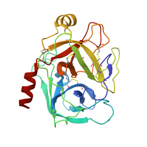
3VPK

Crystal Structure of 6-Guanidinohexanoyl Trypsin


Experimental Data Snapshot

  • Method: X-RAY DIFFRACTION
  • Resolution: 1.94 Å
  • R-Value Free: 0.217 
  • R-Value Work: 0.185 
  • R-Value Observed: 0.186 

wwPDB Validation   3D Report Full Report


Ligand Structure Quality Assessment 


This is version 1.1 of the entry. See complete history


Literature

Crystal structure of 6-guanidinohexanoyl trypsin near the optimum pH reveals the acyl-enzyme intermediate to be deacylated

Masuda, Y.Nitanai, Y.Mizutani, R.Noguchi, S.

(2012) Proteins 

  • DOI: https://doi.org/10.1002/prot.24206
  • Primary Citation of Related Structures:  
    3VPK

  • PubMed Abstract: 

    The force driving the conversion from the acyl intermediate to the tetrahedral intermediate in the deacylation reaction of serine proteases remains unclear. The crystal structure of 6-guanidinohexanoyl trypsin was determined at pH 7.0, near the optimum reaction pH, at 1.94 Å resolution. In this structure, three water molecules are observed around the catalytic site. One acts as a nucleophile to attack the acyl carbonyl carbon while the other two waters fix the position of the catalytic water through a hydrogen bond. When the acyl carbonyl oxygen oscillates thermally, the water assumes an appropriate angle to catalyze the deacylation.


  • Organizational Affiliation

    Nuclear, Biological and Chemical Detection Technology Section, Human Oriented Systems Division, Advanced Defense Technology Center, Technical Research and Development Institute, Ministry of Defense, Meguro, Tokyo 153-8630, Japan. mas@cs.trdi.mod.go.jp


Macromolecules
Find similar proteins by:  (by identity cutoff)  |  3D Structure
Entity ID: 1
MoleculeChains Sequence LengthOrganismDetailsImage
Cationic trypsin223Bos taurusMutation(s): 0 
EC: 3.4.21.4
UniProt
Find proteins for P00760 (Bos taurus)
Explore P00760 
Go to UniProtKB:  P00760
Entity Groups  
Sequence Clusters30% Identity50% Identity70% Identity90% Identity95% Identity100% Identity
UniProt GroupP00760
Sequence Annotations
Expand
  • Reference Sequence
Experimental Data & Validation

Experimental Data

  • Method: X-RAY DIFFRACTION
  • Resolution: 1.94 Å
  • R-Value Free: 0.217 
  • R-Value Work: 0.185 
  • R-Value Observed: 0.186 
  • Space Group: P 21 21 21
Unit Cell:
Length ( Å )Angle ( ˚ )
a = 63.779α = 90
b = 61.05β = 90
c = 69.664γ = 90
Software Package:
Software NamePurpose
SCALEPACKdata scaling
REFMACrefinement
PDB_EXTRACTdata extraction
DENZOdata reduction
SOLVEphasing

Structure Validation

View Full Validation Report


Ligand Structure Quality Assessment 


Entry History 

Deposition Data

Revision History  (Full details and data files)

  • Version 1.0: 2012-05-23
    Type: Initial release
  • Version 1.1: 2013-02-06
    Changes: Database references