6VMS

Structure of a D2 dopamine receptor-G-protein complex in a lipid membrane


Experimental Data Snapshot

  • Method: ELECTRON MICROSCOPY
  • Resolution: 3.80 Å
  • Aggregation State: PARTICLE 
  • Reconstruction Method: SINGLE PARTICLE 

wwPDB Validation   3D Report Full Report


This is version 1.2 of the entry. See complete history


Literature

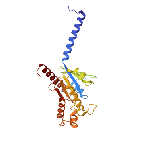
Structure of a D2 dopamine receptor-G-protein complex in a lipid membrane.

Yin, J.Chen, K.M.Clark, M.J.Hijazi, M.Kumari, P.Bai, X.C.Sunahara, R.K.Barth, P.Rosenbaum, D.M.

(2020) Nature 584: 125-129

  • DOI: https://doi.org/10.1038/s41586-020-2379-5
  • Primary Citation of Related Structures:  
    6VMS

  • PubMed Abstract: 

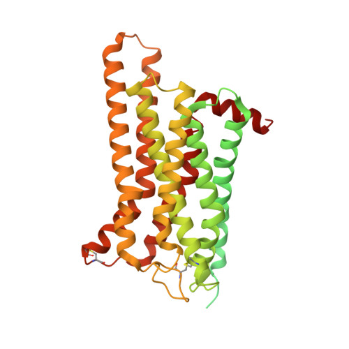
    The D2 dopamine receptor (DRD2) is a therapeutic target for Parkinson's disease 1 and antipsychotic drugs 2 . DRD2 is activated by the endogenous neurotransmitter dopamine and synthetic agonist drugs such as bromocriptine 3 , leading to stimulation of G i and inhibition of adenylyl cyclase. Here we used cryo-electron microscopy to elucidate the structure of an agonist-bound activated DRD2-G i complex reconstituted into a phospholipid membrane. The extracellular ligand-binding site of DRD2 is remodelled in response to agonist binding, with conformational changes in extracellular loop 2, transmembrane domain 5 (TM5), TM6 and TM7, propagating to opening of the intracellular G i -binding site. The DRD2-G i structure represents, to our knowledge, the first experimental model of a G-protein-coupled receptor-G-protein complex embedded in a phospholipid bilayer, which serves as a benchmark to validate the interactions seen in previous detergent-bound structures. The structure also reveals interactions that are unique to the membrane-embedded complex, including helix 8 burial in the inner leaflet, ordered lysine and arginine side chains in the membrane interfacial regions, and lipid anchoring of the G protein in the membrane. Our model of the activated DRD2 will help to inform the design of subtype-selective DRD2 ligands for multiple human central nervous system disorders.


  • Organizational Affiliation

    Department of Biophysics, The University of Texas Southwestern Medical Center, Dallas, TX, USA.


Macromolecules
Find similar proteins by:  (by identity cutoff)  |  3D Structure
Entity ID: 1
MoleculeChains Sequence LengthOrganismDetailsImage
Guanine nucleotide-binding protein G(i) subunit alpha-1354Rattus norvegicusMutation(s): 0 
Gene Names: Gnai1Gnai-1
Membrane Entity: Yes 
UniProt
Find proteins for P10824 (Rattus norvegicus)
Explore P10824 
Go to UniProtKB:  P10824
Entity Groups  
Sequence Clusters30% Identity50% Identity70% Identity90% Identity95% Identity100% Identity
UniProt GroupP10824
Sequence Annotations
Expand
  • Reference Sequence
Find similar proteins by:  (by identity cutoff)  |  3D Structure
Entity ID: 2
MoleculeChains Sequence LengthOrganismDetailsImage
Guanine nucleotide-binding protein G(I)/G(S)/G(T) subunit beta-1340Homo sapiensMutation(s): 0 
Gene Names: GNB1
UniProt & NIH Common Fund Data Resources
Find proteins for P62873 (Homo sapiens)
Explore P62873 
Go to UniProtKB:  P62873
PHAROS:  P62873
GTEx:  ENSG00000078369 
Entity Groups  
Sequence Clusters30% Identity50% Identity70% Identity90% Identity95% Identity100% Identity
UniProt GroupP62873
Sequence Annotations
Expand
  • Reference Sequence
Find similar proteins by:  (by identity cutoff)  |  3D Structure
Entity ID: 3
MoleculeChains Sequence LengthOrganismDetailsImage
Guanine nucleotide-binding protein G(I)/G(S)/G(O) subunit gamma-282Homo sapiensMutation(s): 0 
Gene Names: GNG2
Membrane Entity: Yes 
UniProt & NIH Common Fund Data Resources
Find proteins for P59768 (Homo sapiens)
Explore P59768 
Go to UniProtKB:  P59768
PHAROS:  P59768
GTEx:  ENSG00000186469 
Entity Groups  
Sequence Clusters30% Identity50% Identity70% Identity90% Identity95% Identity100% Identity
UniProt GroupP59768
Sequence Annotations
Expand
  • Reference Sequence
Find similar proteins by:  (by identity cutoff)  |  3D Structure
Entity ID: 4
MoleculeChains Sequence LengthOrganismDetailsImage
scFv16D [auth E]259Homo sapiensMutation(s): 0 
Entity Groups  
Sequence Clusters30% Identity50% Identity70% Identity90% Identity95% Identity100% Identity
Sequence Annotations
Expand
  • Reference Sequence
Find similar proteins by:  (by identity cutoff)  |  3D Structure
Entity ID: 5
MoleculeChains Sequence LengthOrganismDetailsImage
Endolysin,D(2) dopamine receptor,D(2) dopamine receptorE [auth R]450Tequatrovirus T4Homo sapiensMutation(s): 0 
Gene Names: eT4Tp126DRD2
EC: 3.2.1.17
Membrane Entity: Yes 
UniProt & NIH Common Fund Data Resources
Find proteins for P14416 (Homo sapiens)
Explore P14416 
Go to UniProtKB:  P14416
PHAROS:  P14416
GTEx:  ENSG00000149295 
Find proteins for D9IEF7 (Enterobacteria phage T4)
Explore D9IEF7 
Go to UniProtKB:  D9IEF7
Entity Groups  
Sequence Clusters30% Identity50% Identity70% Identity90% Identity95% Identity100% Identity
UniProt GroupsP14416D9IEF7
Sequence Annotations
Expand
  • Reference Sequence
Small Molecules
Ligands 1 Unique
IDChains Name / Formula / InChI Key2D Diagram3D Interactions
08Y (Subject of Investigation/LOI)
Query on 08Y

Download Ideal Coordinates CCD File 
F [auth R]bromoergocryptine
C32 H40 Br N5 O5
OZVBMTJYIDMWIL-AYFBDAFISA-N
Experimental Data & Validation

Experimental Data

  • Method: ELECTRON MICROSCOPY
  • Resolution: 3.80 Å
  • Aggregation State: PARTICLE 
  • Reconstruction Method: SINGLE PARTICLE 
EM Software:
TaskSoftware PackageVersion
RECONSTRUCTIONRELION3.0.1
MODEL REFINEMENTPHENIX

Structure Validation

View Full Validation Report


Entry History & Funding Information

Deposition Data


Funding OrganizationLocationGrant Number
Welch FoundationUnited StatesI-1770

Revision History  (Full details and data files)

  • Version 1.0: 2020-06-17
    Type: Initial release
  • Version 1.1: 2020-06-24
    Changes: Database references
  • Version 1.2: 2020-08-19
    Changes: Database references