2AEY

Crystal structure of fructan 1-exohydrolase IIa from Cichorium intybus in complex with 2,5 dideoxy-2,5-immino-D-mannitol


Experimental Data Snapshot

  • Method: X-RAY DIFFRACTION
  • Resolution: 3.27 Å
  • R-Value Free: 0.257 
  • R-Value Work: 0.217 
  • R-Value Observed: 0.217 

wwPDB Validation   3D Report Full Report


Ligand Structure Quality Assessment 


This is version 2.1 of the entry. See complete history


Literature

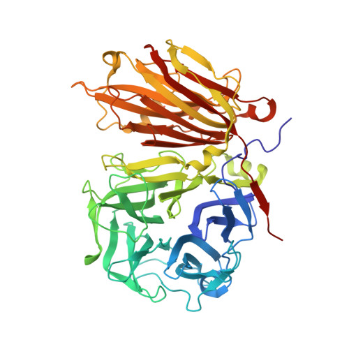
Insights into the fine architecture of the active site of chicory fructan 1-exohydrolase: 1-kestose as substrate vs sucrose as inhibitor.

Verhaest, M.Lammens, W.Le Roy, K.De Ranter, C.J.Van Laere, A.Rabijns, A.Van den Ende, W.

(2007) New Phytol 174: 90-100

  • DOI: https://doi.org/10.1111/j.1469-8137.2007.01988.x
  • Primary Citation of Related Structures:  
    2ADD, 2ADE, 2AEY, 2AEZ

  • PubMed Abstract: 

    * Invertases and fructan exohydrolases (FEHs) fulfil important physiological functions in plants. Sucrose is the typical substrate for invertases and bacterial levansucrases but not for plant FEHs, which are usually inhibited by sucrose. * Here we report on complexes between chicory (Cichorium intybus) 1-FEH IIa with the substrate 1-kestose and the inhibitors sucrose, fructose and 2,5 dideoxy-2,5-imino-D-mannitol. Comparisons with other family GH32 and 68 enzyme-substrate complexes revealed that sucrose can bind as a substrate (invertase/levansucrase) or as an inhibitor (1-FEH IIa). * Sucrose acts as inhibitor because the O2 of the glucose moiety forms an H-linkage with the acid-base catalyst E201, inhibiting catalysis. By contrast, the homologous O3 of the internal fructose in the substrate 1-kestose forms an intramolecular H-linkage and does not interfere with the catalytic process. Mutagenesis showed that W82 and S101 are important for binding sucrose as inhibitor. * The physiological implications of the essential differences in the active sites of FEHs and invertases/levansucrases are discussed. Sucrose-inhibited FEHs show a K(i) (inhibition constant) well below physiological sucrose concentrations and could be rapidly activated under carbon deprivation.


  • Organizational Affiliation

    Laboratorium voor Biokristallografie, Faculteit Farmaceutische Wetenschappen, K.U. Leuven, Herestraat 49, O & N II, bus 822, B-3000 Leuven, Belgium.


Macromolecules
Find similar proteins by:  (by identity cutoff)  |  3D Structure
Entity ID: 1
MoleculeChains Sequence LengthOrganismDetailsImage
fructan 1-exohydrolase IIa543Cichorium intybusMutation(s): 0 
Gene Names: 1-feh IIa
EC: 3.2.1.153
UniProt
Find proteins for Q93X60 (Cichorium intybus)
Explore Q93X60 
Go to UniProtKB:  Q93X60
Entity Groups  
Sequence Clusters30% Identity50% Identity70% Identity90% Identity95% Identity100% Identity
UniProt GroupQ93X60
Sequence Annotations
Expand
  • Reference Sequence
Oligosaccharides

Help

Entity ID: 2
MoleculeChains Length2D Diagram Glycosylation3D Interactions
alpha-D-mannopyranose-(1-4)-2-acetamido-2-deoxy-beta-D-glucopyranose-(1-4)-2-acetamido-2-deoxy-beta-D-glucopyranose
B
3N-Glycosylation
Glycosylation Resources
GlyTouCan:  G62182OO
GlyCosmos:  G62182OO
GlyGen:  G62182OO
Entity ID: 3
MoleculeChains Length2D Diagram Glycosylation3D Interactions
2-acetamido-2-deoxy-beta-D-glucopyranose-(1-4)-2-acetamido-2-deoxy-beta-D-glucopyranose
C
2N-Glycosylation
Glycosylation Resources
GlyTouCan:  G42666HT
GlyCosmos:  G42666HT
GlyGen:  G42666HT
Small Molecules
Ligands 1 Unique
IDChains Name / Formula / InChI Key2D Diagram3D Interactions
DQQ
Query on DQQ

Download Ideal Coordinates CCD File 
D [auth A]2,5-DIDEOXY-2,5-IMINO-D-MANNITOL
C6 H13 N O4
PFYHYHZGDNWFIF-KVTDHHQDSA-N
Experimental Data & Validation

Experimental Data

  • Method: X-RAY DIFFRACTION
  • Resolution: 3.27 Å
  • R-Value Free: 0.257 
  • R-Value Work: 0.217 
  • R-Value Observed: 0.217 
  • Space Group: P 41 21 2
Unit Cell:
Length ( Å )Angle ( ˚ )
a = 138.86α = 90
b = 138.86β = 90
c = 182.44γ = 90
Software Package:
Software NamePurpose
CNSrefinement
DENZOdata reduction
SCALEPACKdata scaling
CNSphasing

Structure Validation

View Full Validation Report


Ligand Structure Quality Assessment 


Entry History 

Deposition Data

Revision History  (Full details and data files)

  • Version 1.0: 2006-08-29
    Type: Initial release
  • Version 1.1: 2008-04-30
    Changes: Version format compliance
  • Version 1.2: 2011-07-13
    Changes: Non-polymer description, Version format compliance
  • Version 2.0: 2020-07-29
    Type: Remediation
    Reason: Carbohydrate remediation
    Changes: Advisory, Atomic model, Data collection, Derived calculations, Structure summary
  • Version 2.1: 2023-08-23
    Changes: Data collection, Database references, Refinement description, Structure summary