3MJG

The structure of a platelet derived growth factor receptor complex


Experimental Data Snapshot

  • Method: X-RAY DIFFRACTION
  • Resolution: 2.30 Å
  • R-Value Free: 0.277 
  • R-Value Work: 0.237 
  • R-Value Observed: 0.237 

wwPDB Validation   3D Report Full Report


Ligand Structure Quality Assessment 


This is version 2.1 of the entry. See complete history


Literature

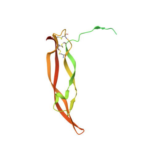
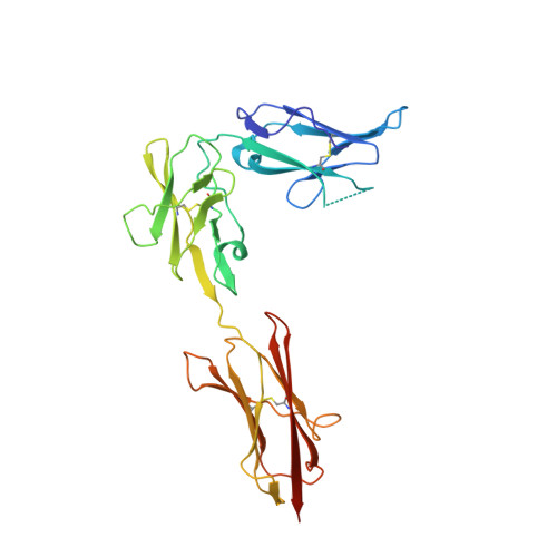
Structures of a platelet-derived growth factor/propeptide complex and a platelet-derived growth factor/receptor complex.

Shim, A.H.Liu, H.Focia, P.J.Chen, X.Lin, P.C.He, X.

(2010) Proc Natl Acad Sci U S A 107: 11307-11312

  • DOI: https://doi.org/10.1073/pnas.1000806107
  • Primary Citation of Related Structures:  
    3MJG, 3MJK

  • PubMed Abstract: 

    Platelet-derived growth factors (PDGFs) and their receptors (PDGFRs) are prototypic growth factors and receptor tyrosine kinases which have critical functions in development. We show that PDGFs share a conserved region in their prodomain sequences which can remain noncovalently associated with the mature cystine-knot growth factor domain after processing. The structure of the PDGF-A/propeptide complex reveals this conserved, hydrophobic association mode. We also present the structure of the complex between PDGF-B and the first three Ig domains of PDGFRbeta, showing that two PDGF-B protomers clamp PDGFRbeta at their dimerization seam. The PDGF-B:PDGFRbeta interface is predominantly hydrophobic, and PDGFRs and the PDGF propeptides occupy overlapping positions on mature PDGFs, rationalizing the need of propeptides by PDGFs to cover functionally important hydrophobic surfaces during secretion. A large-scale structural organization and rearrangement is observed for PDGF-B upon receptor binding, in which the PDGF-B L1 loop, disordered in the structure of the free form, adopts a highly specific conformation to form hydrophobic interactions with the third Ig domain of PDGFRbeta. Calorimetric data also shows that the membrane-proximal homotypic PDGFRalpha interaction, albeit required for activation, contributes negatively to ligand binding. The structural and biochemical data together offer insights into PDGF-PDGFR signaling, as well as strategies for PDGF-antagonism.


  • Organizational Affiliation

    Department of Molecular Pharmacology and Biological Chemistry, Northwestern University Feinberg School of Medicine, Searle 8-417, 303 East Chicago Avenue, Chicago, IL 60611, USA.


Macromolecules
Find similar proteins by:  (by identity cutoff)  |  3D Structure
Entity ID: 1
MoleculeChains Sequence LengthOrganismDetailsImage
Platelet-derived growth factor subunit B
A, B
172Homo sapiensMutation(s): 0 
Gene Names: PDGF2PDGFBSIS
UniProt & NIH Common Fund Data Resources
Find proteins for P01127 (Homo sapiens)
Explore P01127 
Go to UniProtKB:  P01127
PHAROS:  P01127
GTEx:  ENSG00000100311 
Entity Groups  
Sequence Clusters30% Identity50% Identity70% Identity90% Identity95% Identity100% Identity
UniProt GroupP01127
Sequence Annotations
Expand
  • Reference Sequence
Find similar proteins by:  (by identity cutoff)  |  3D Structure
Entity ID: 2
MoleculeChains Sequence LengthOrganismDetailsImage
Beta-type platelet-derived growth factor receptorC [auth X],
D [auth Y]
289Homo sapiensMutation(s): 0 
Gene Names: PDGFRB
EC: 2.7.10.1
UniProt & NIH Common Fund Data Resources
Find proteins for P09619 (Homo sapiens)
Explore P09619 
Go to UniProtKB:  P09619
PHAROS:  P09619
GTEx:  ENSG00000113721 
Entity Groups  
Sequence Clusters30% Identity50% Identity70% Identity90% Identity95% Identity100% Identity
UniProt GroupP09619
Sequence Annotations
Expand
  • Reference Sequence
Small Molecules
Ligands 2 Unique
IDChains Name / Formula / InChI Key2D Diagram3D Interactions
NAG
Query on NAG

Download Ideal Coordinates CCD File 
F [auth X]
G [auth X]
H [auth X]
I [auth X]
J [auth X]
F [auth X],
G [auth X],
H [auth X],
I [auth X],
J [auth X],
K [auth X],
L [auth Y],
M [auth Y],
N [auth Y],
O [auth Y],
P [auth Y],
Q [auth Y],
R [auth Y]
2-acetamido-2-deoxy-beta-D-glucopyranose
C8 H15 N O6
OVRNDRQMDRJTHS-FMDGEEDCSA-N
NDG
Query on NDG

Download Ideal Coordinates CCD File 
E [auth X]2-acetamido-2-deoxy-alpha-D-glucopyranose
C8 H15 N O6
OVRNDRQMDRJTHS-PVFLNQBWSA-N
Experimental Data & Validation

Experimental Data

  • Method: X-RAY DIFFRACTION
  • Resolution: 2.30 Å
  • R-Value Free: 0.277 
  • R-Value Work: 0.237 
  • R-Value Observed: 0.237 
  • Space Group: P 21 21 21
Unit Cell:
Length ( Å )Angle ( ˚ )
a = 78.47α = 90
b = 116.82β = 90
c = 134.15γ = 90
Software Package:
Software NamePurpose
HKL-2000data collection
PHASERphasing
CNSrefinement
HKL-2000data reduction
HKL-2000data scaling

Structure Validation

View Full Validation Report


Ligand Structure Quality Assessment 


Entry History 

Deposition Data

Revision History  (Full details and data files)

  • Version 1.0: 2010-06-16
    Type: Initial release
  • Version 1.1: 2011-07-13
    Changes: Version format compliance
  • Version 1.2: 2017-11-08
    Changes: Source and taxonomy
  • Version 2.0: 2020-07-29
    Type: Remediation
    Reason: Carbohydrate remediation
    Changes: Advisory, Atomic model, Data collection, Database references, Derived calculations, Structure summary
  • Version 2.1: 2023-09-06
    Changes: Data collection, Database references, Refinement description, Structure summary