6BTH

Crystal structure of human cellular retinol binding protein 2 (CRBP2) in complex with 2-arachidonoylglycerol (2-AG)


Experimental Data Snapshot

  • Method: X-RAY DIFFRACTION
  • Resolution: 1.35 Å
  • R-Value Free: 0.164 
  • R-Value Work: 0.126 
  • R-Value Observed: 0.128 

wwPDB Validation   3D Report Full Report


Ligand Structure Quality Assessment 


This is version 1.3 of the entry. See complete history


Literature

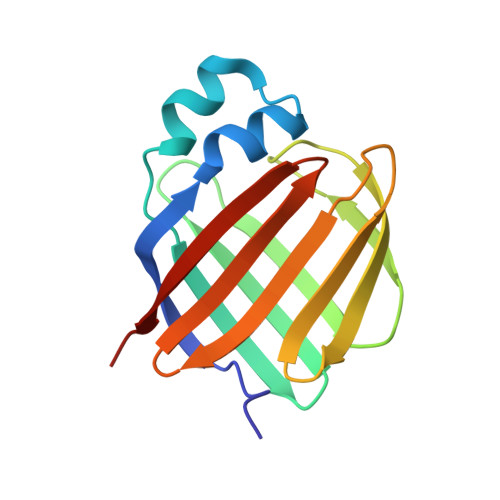
Retinol-binding protein 2 (RBP2) binds monoacylglycerols and modulates gut endocrine signaling and body weight.

Lee, S.A.Yang, K.J.Z.Brun, P.J.Silvaroli, J.A.Yuen, J.J.Shmarakov, I.Jiang, H.Feranil, J.B.Li, X.Lackey, A.I.Krezel, W.Leibel, R.L.Libien, J.Storch, J.Golczak, M.Blaner, W.S.

(2020) Sci Adv 6: eaay8937-eaay8937

  • DOI: https://doi.org/10.1126/sciadv.aay8937
  • Primary Citation of Related Structures:  
    6BTH, 6BTI

  • PubMed Abstract: 

    Expressed in the small intestine, retinol-binding protein 2 (RBP2) facilitates dietary retinoid absorption. Rbp2 -deficient ( Rbp2 -/- ) mice fed a chow diet exhibit by 6-7 months-of-age higher body weights, impaired glucose metabolism, and greater hepatic triglyceride levels compared to controls. These phenotypes are also observed when young Rbp2 -/- mice are fed a high fat diet. Retinoids do not account for the phenotypes. Rather, RBP2 is a previously unidentified monoacylglycerol (MAG)-binding protein, interacting with the endocannabinoid 2-arachidonoylglycerol (2-AG) and other MAGs with affinities comparable to retinol. X-ray crystallographic studies show that MAGs bind in the retinol binding pocket. When challenged with an oil gavage, Rbp2 -/- mice show elevated mucosal levels of 2-MAGs. This is accompanied by significantly elevated blood levels of the gut hormone GIP (glucose-dependent insulinotropic polypeptide). Thus, RBP2, in addition to facilitating dietary retinoid absorption, modulates MAG metabolism and likely signaling, playing a heretofore unknown role in systemic energy balance.


  • Organizational Affiliation

    Department of Medicine, Institute of Human Nutrition, College of Physicians and Surgeons, Columbia University, New York, NY, USA.


Macromolecules
Find similar proteins by:  (by identity cutoff)  |  3D Structure
Entity ID: 1
MoleculeChains Sequence LengthOrganismDetailsImage
Retinol-binding protein 2
A, B
138Homo sapiensMutation(s): 0 
Gene Names: RBP2CRBP2
UniProt & NIH Common Fund Data Resources
Find proteins for P50120 (Homo sapiens)
Explore P50120 
Go to UniProtKB:  P50120
PHAROS:  P50120
GTEx:  ENSG00000114113 
Entity Groups  
Sequence Clusters30% Identity50% Identity70% Identity90% Identity95% Identity100% Identity
UniProt GroupP50120
Sequence Annotations
Expand
  • Reference Sequence
Experimental Data & Validation

Experimental Data

  • Method: X-RAY DIFFRACTION
  • Resolution: 1.35 Å
  • R-Value Free: 0.164 
  • R-Value Work: 0.126 
  • R-Value Observed: 0.128 
  • Space Group: P 21 21 21
Unit Cell:
Length ( Å )Angle ( ˚ )
a = 58.138α = 90
b = 67.328β = 90
c = 87.319γ = 90
Software Package:
Software NamePurpose
PHENIXrefinement
XDSdata reduction
SCALAdata scaling
PHASERphasing

Structure Validation

View Full Validation Report


Ligand Structure Quality Assessment 


Entry History & Funding Information

Deposition Data


Funding OrganizationLocationGrant Number
National Institutes of Health/National Eye Institute (NIH/NEI)United StatesR01-EY023948

Revision History  (Full details and data files)

  • Version 1.0: 2018-12-12
    Type: Initial release
  • Version 1.1: 2019-12-18
    Changes: Author supporting evidence
  • Version 1.2: 2020-05-20
    Changes: Database references
  • Version 1.3: 2023-10-04
    Changes: Data collection, Database references, Refinement description