6M13

Crystal structure of Rnase L in complex with Toceranib


Experimental Data Snapshot

  • Method: X-RAY DIFFRACTION
  • Resolution: 2.56 Å
  • R-Value Free: 0.286 
  • R-Value Work: 0.231 
  • R-Value Observed: 0.234 

wwPDB Validation   3D Report Full Report


Ligand Structure Quality Assessment 


This is version 1.2 of the entry. See complete history


Literature

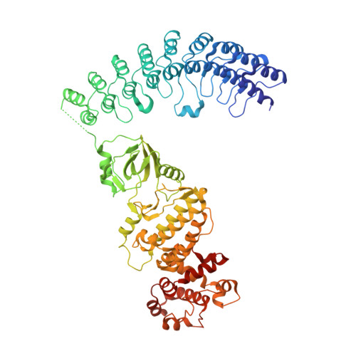
Sunitinib inhibits RNase L by destabilizing its active dimer conformation.

Tang, J.Wang, Y.Zhou, H.Ye, Y.Talukdar, M.Fu, Z.Liu, Z.Li, J.Neculai, D.Gao, J.Huang, H.

(2020) Biochem J 477: 3387-3399

  • DOI: https://doi.org/10.1042/BCJ20200260
  • Primary Citation of Related Structures:  
    6M11, 6M12, 6M13

  • PubMed Abstract: 

    The pseudokinase (PK) RNase L is a functional ribonuclease and plays important roles in human innate immunity. The ribonuclease activity of RNase L can be regulated by the kinase inhibitor sunitinib. The combined use of oncolytic virus and sunitinib has been shown to exert synergistic effects in anticancer therapy. In this study, we aimed to uncover the mechanism of action through which sunitinib inhibits RNase L. We solved the crystal structures of RNase L in complex with sunitinib and its analogs toceranib and SU11652. Our results showed that sunitinib bound to the ATP-binding pocket of RNase L. Unexpectedly, the αA helix linking the ankyrin repeat-domain and the PK domain affected the binding mode of sunitinib and resulted in an unusual flipped orientation relative to other structures in PDB. Molecular dynamics simulations and dynamic light scattering results support that the binding of sunitinib in the PK domain destabilized the dimer conformation of RNase L and allosterically inhibited its ribonuclease activity. Our study suggested that dimer destabilization could be an effective strategy for the discovery of RNase L inhibitors and that targeting the ATP-binding pocket in the PK domain of RNase L was an efficient approach for modulating its ribonuclease activity.


  • Organizational Affiliation

    State Key Laboratory of Chemical Oncogenomics, School of Chemical Biology and Biotechnology, Peking University Shenzhen Graduate School, Shenzhen, China.


Macromolecules
Find similar proteins by:  (by identity cutoff)  |  3D Structure
Entity ID: 1
MoleculeChains Sequence LengthOrganismDetailsImage
Ribonuclease LA [auth a],
B [auth b]
717Sus scrofaMutation(s): 0 
Gene Names: RNASEL
UniProt
Find proteins for A5H025 (Sus scrofa)
Explore A5H025 
Go to UniProtKB:  A5H025
Entity Groups  
Sequence Clusters30% Identity50% Identity70% Identity90% Identity95% Identity100% Identity
UniProt GroupA5H025
Sequence Annotations
Expand
  • Reference Sequence
Small Molecules
Ligands 3 Unique
IDChains Name / Formula / InChI Key2D Diagram3D Interactions
25L
Query on 25L

Download Ideal Coordinates CCD File 
D [auth a],
G [auth b]
[[(2R,3R,4R,5R)-5-(6-aminopurin-9-yl)-4-[[(2R,3R,4R,5R)-5-(6-aminopurin-9-yl)-4-[[(2R,3S,4R,5R)-5-(6-aminopurin-9-yl)-3,4-dihydroxy-oxolan-2-yl]methoxy-hydroxy-phosphoryl]oxy-3-hydroxy-oxolan-2-yl]methoxy-hydroxy-phosphoryl]oxy-3-hydroxy-oxolan-2-yl]methoxy-hydroxy-phosphoryl] phosphono hydrogen phosphate
C30 H40 N15 O25 P5
RTAGLZBJCCVJET-UQTMIEBXSA-N
BWC (Subject of Investigation/LOI)
Query on BWC

Download Ideal Coordinates CCD File 
C [auth a],
F [auth b]
5-[(Z)-(5-fluoranyl-2-oxidanylidene-1H-indol-3-ylidene)methyl]-2,4-dimethyl-N-(2-pyrrolidin-1-ylethyl)-1H-pyrrole-3-carboxamide
C22 H25 F N4 O2
SRSGVKWWVXWSJT-ATVHPVEESA-N
PO4
Query on PO4

Download Ideal Coordinates CCD File 
E [auth a]PHOSPHATE ION
O4 P
NBIIXXVUZAFLBC-UHFFFAOYSA-K
Experimental Data & Validation

Experimental Data

  • Method: X-RAY DIFFRACTION
  • Resolution: 2.56 Å
  • R-Value Free: 0.286 
  • R-Value Work: 0.231 
  • R-Value Observed: 0.234 
  • Space Group: P 21 21 21
Unit Cell:
Length ( Å )Angle ( ˚ )
a = 59.45α = 90
b = 111.36β = 90
c = 263.86γ = 90
Software Package:
Software NamePurpose
REFMACrefinement
xia2data scaling
PDB_EXTRACTdata extraction
XDSdata reduction
PHASERphasing

Structure Validation

View Full Validation Report


Ligand Structure Quality Assessment 


Entry History & Funding Information

Deposition Data


Funding OrganizationLocationGrant Number
National Science Foundation (NSF, China)China--

Revision History  (Full details and data files)

  • Version 1.0: 2020-09-02
    Type: Initial release
  • Version 1.1: 2020-09-30
    Changes: Database references
  • Version 1.2: 2023-11-29
    Changes: Data collection, Database references, Refinement description