5D7D

Crystal structure of the ATP binding domain of S. aureus GyrB complexed with a ligand


Experimental Data Snapshot

  • Method: X-RAY DIFFRACTION
  • Resolution: 1.60 Å
  • R-Value Free: 0.195 
  • R-Value Work: 0.165 
  • R-Value Observed: 0.167 

wwPDB Validation   3D Report Full Report


Ligand Structure Quality Assessment 


This is version 1.2 of the entry. See complete history


Literature

Discovery of Indazole Derivatives as a Novel Class of Bacterial Gyrase B Inhibitors.

Zhang, J.Yang, Q.Romero, J.A.Cross, J.Wang, B.Poutsiaka, K.M.Epie, F.Bevan, D.Wu, Y.Moy, T.Daniel, A.Chamberlain, B.Carter, N.Shotwell, J.Arya, A.Kumar, V.Silverman, J.Nguyen, K.Metcalf, C.A.Ryan, D.Lippa, B.Dolle, R.E.

(2015) ACS Med Chem Lett 6: 1080-1085

  • DOI: https://doi.org/10.1021/acsmedchemlett.5b00266
  • Primary Citation of Related Structures:  
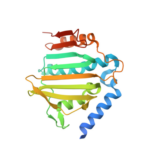
    5D7D, 5D7R

  • PubMed Abstract: 

    Antibacterials with a novel mechanism of action offer a great opportunity to combat widespread antimicrobial resistance. Bacterial DNA Gyrase is a clinically validated target. Through physiochemical property optimization of a pyrazolopyridone hit, a novel class of GyrB inhibitors were discovered. Guided by structure-based drug design, indazole derivatives with excellent enzymatic and antibacterial activity as well as great animal efficacy were discovered.


  • Organizational Affiliation

    Cubist Pharmaceuticals Inc. , Lexington, Massachusetts 02421, United States.


Macromolecules
Find similar proteins by:  (by identity cutoff)  |  3D Structure
Entity ID: 1
MoleculeChains Sequence LengthOrganismDetailsImage
DNA gyrase subunit B
A, B
212Staphylococcus aureusMutation(s): 0 
Gene Names: gyrB
EC: 5.99.1.3
UniProt
Find proteins for P0A0K8 (Staphylococcus aureus)
Explore P0A0K8 
Go to UniProtKB:  P0A0K8
Entity Groups  
Sequence Clusters30% Identity50% Identity70% Identity90% Identity95% Identity100% Identity
UniProt GroupP0A0K8
Sequence Annotations
Expand
  • Reference Sequence
Small Molecules
Ligands 4 Unique
IDChains Name / Formula / InChI Key2D Diagram3D Interactions
57X
Query on 57X

Download Ideal Coordinates CCD File 
I [auth A],
Q [auth B]
7-propyl-3-[2-(pyridin-3-yl)-1,3-thiazol-5-yl]-1,7-dihydro-6H-pyrazolo[3,4-b]pyridin-6-one
C17 H15 N5 O S
VOOCDNWLHBWIJZ-UHFFFAOYSA-N
MPD
Query on MPD

Download Ideal Coordinates CCD File 
C [auth A],
D [auth A],
K [auth B],
L [auth B]
(4S)-2-METHYL-2,4-PENTANEDIOL
C6 H14 O2
SVTBMSDMJJWYQN-YFKPBYRVSA-N
CL
Query on CL

Download Ideal Coordinates CCD File 
E [auth A],
M [auth B]
CHLORIDE ION
Cl
VEXZGXHMUGYJMC-UHFFFAOYSA-M
MG
Query on MG

Download Ideal Coordinates CCD File 
F [auth A]
G [auth A]
H [auth A]
J [auth A]
N [auth B]
F [auth A],
G [auth A],
H [auth A],
J [auth A],
N [auth B],
O [auth B],
P [auth B]
MAGNESIUM ION
Mg
JLVVSXFLKOJNIY-UHFFFAOYSA-N
Binding Affinity Annotations 
IDSourceBinding Affinity
57X BindingDB:  5D7D IC50: 6 (nM) from 1 assay(s)
Experimental Data & Validation

Experimental Data

  • Method: X-RAY DIFFRACTION
  • Resolution: 1.60 Å
  • R-Value Free: 0.195 
  • R-Value Work: 0.165 
  • R-Value Observed: 0.167 
  • Space Group: C 1 2 1
Unit Cell:
Length ( Å )Angle ( ˚ )
a = 143.3α = 90
b = 55.446β = 100.95
c = 51.029γ = 90
Software Package:
Software NamePurpose
REFMACrefinement
d*TREKdata scaling
MOLREPphasing
PDB_EXTRACTdata extraction
d*TREKdata reduction

Structure Validation

View Full Validation Report


Ligand Structure Quality Assessment 


Entry History 

Revision History  (Full details and data files)

  • Version 1.0: 2015-11-11
    Type: Initial release
  • Version 1.1: 2017-11-22
    Changes: Derived calculations, Refinement description
  • Version 1.2: 2023-09-27
    Changes: Data collection, Database references, Derived calculations, Refinement description