8OOG

Crystal structure of human MAT2a with S-Adenosylmethionine and a fragment bound in a novel pocket


Experimental Data Snapshot

  • Method: X-RAY DIFFRACTION
  • Resolution: 1.38 Å
  • R-Value Free: 0.200 
  • R-Value Work: 0.190 
  • R-Value Observed: 0.191 

wwPDB Validation   3D Report Full Report


Ligand Structure Quality Assessment 


This is version 1.0 of the entry. See complete history


Literature

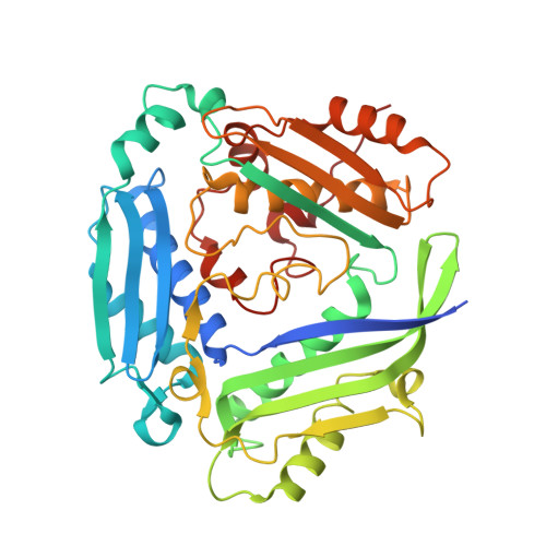
Combining structural and coevolution information to unveil allosteric sites.

La Sala, G.Pfleger, C.Kack, H.Wissler, L.Nevin, P.Bohm, K.Janet, J.P.Schimpl, M.Stubbs, C.J.De Vivo, M.Tyrchan, C.Hogner, A.Gohlke, H.Frolov, A.I.

(2023) Chem Sci 14: 7057-7067

  • DOI: https://doi.org/10.1039/d2sc06272k
  • Primary Citation of Related Structures:  
    8OOG

  • PubMed Abstract: 

    Understanding allosteric regulation in biomolecules is of great interest to pharmaceutical research and computational methods emerged during the last decades to characterize allosteric coupling. However, the prediction of allosteric sites in a protein structure remains a challenging task. Here, we integrate local binding site information, coevolutionary information, and information on dynamic allostery into a structure-based three-parameter model to identify potentially hidden allosteric sites in ensembles of protein structures with orthosteric ligands. When tested on five allosteric proteins (LFA-1, p38-α, GR, MAT2A, and BCKDK), the model successfully ranked all known allosteric pockets in the top three positions. Finally, we identified a novel druggable site in MAT2A confirmed by X-ray crystallography and SPR and a hitherto unknown druggable allosteric site in BCKDK validated by biochemical and X-ray crystallography analyses. Our model can be applied in drug discovery to identify allosteric pockets.


  • Organizational Affiliation

    Medicinal Chemistry, Research and Early Development, Cardiovascular, Renal and Metabolism (CVRM), BioPharmaceuticals R&D, AstraZeneca Gothenburg Sweden giuseppina.lasala@astrazeneca.com andrey.frolov@astrazeneca.com.


Macromolecules
Find similar proteins by:  (by identity cutoff)  |  3D Structure
Entity ID: 1
MoleculeChains Sequence LengthOrganismDetailsImage
S-adenosylmethionine synthase isoform type-2397Homo sapiensMutation(s): 0 
Gene Names: MAT2AAMS2MATA2
EC: 2.5.1.6
UniProt & NIH Common Fund Data Resources
Find proteins for P31153 (Homo sapiens)
Explore P31153 
Go to UniProtKB:  P31153
PHAROS:  P31153
GTEx:  ENSG00000168906 
Entity Groups  
Sequence Clusters30% Identity50% Identity70% Identity90% Identity95% Identity100% Identity
UniProt GroupP31153
Sequence Annotations
Expand
  • Reference Sequence
Experimental Data & Validation

Experimental Data

  • Method: X-RAY DIFFRACTION
  • Resolution: 1.38 Å
  • R-Value Free: 0.200 
  • R-Value Work: 0.190 
  • R-Value Observed: 0.191 
  • Space Group: I 2 2 2
Unit Cell:
Length ( Å )Angle ( ˚ )
a = 67.88α = 90
b = 94.22β = 90
c = 116.96γ = 90
Software Package:
Software NamePurpose
MOSFLMdata reduction
SCALAdata scaling
BUSTERrefinement
Aimlessdata scaling
XDSdata reduction
AMoREphasing

Structure Validation

View Full Validation Report


Ligand Structure Quality Assessment 


Entry History & Funding Information

Deposition Data

  • Released Date: 2023-07-12 
  • Deposition Author(s): Schimpl, M.

Funding OrganizationLocationGrant Number
Not funded--

Revision History  (Full details and data files)

  • Version 1.0: 2023-07-12
    Type: Initial release