 Spectrozyme tPA BDBM14168
 tPA/Trypsin Substrate BDBM16177
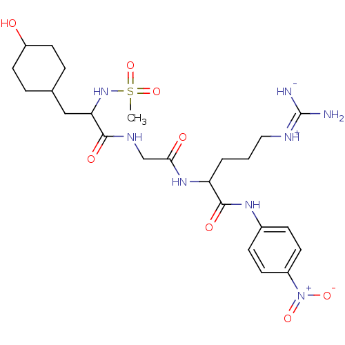 Spectrozyme tPA tPA/Trypsin Substrate N-Methylsulfonyl-D-hexahydrotyrosyl-glycyl-L-arginine-4-nitroanilide acetate Methylsulfonyl-D-hexahydrotyrosyl-glycyl-arginine-4-nitroanilide CH3-SO2-D-HHT-Gly-Arg-pNA BDBM12774 (2S)-5-carbamimidamido-2-{2-[(2R)-3-(4-hydroxycyclohexyl)-2-methanesulfonamidopropanamido]acetamido}-N-(4-nitrophenyl)pentanamide; acetic acid Factor IX Chromogenic Substrate MS-D-HHT-Gly-Arg-pNA
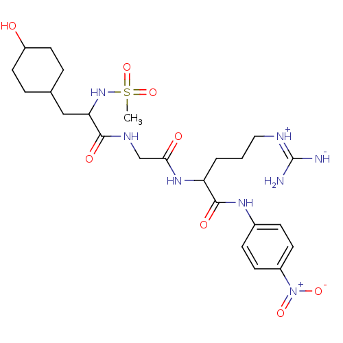 (2S)-5-carbamimidamido-2-{2-[(2R)-3-(4-hydroxycyclohexyl)-2-methanesulfonamidopropanamido]acetamido}-N-(4-nitrophenyl)pentanamide BDBM23868 Pefachrome tPA N-Methylsulfonyl-D-hexahydrotyrosyl-glycyl-L-arginine-4-nitroanilide
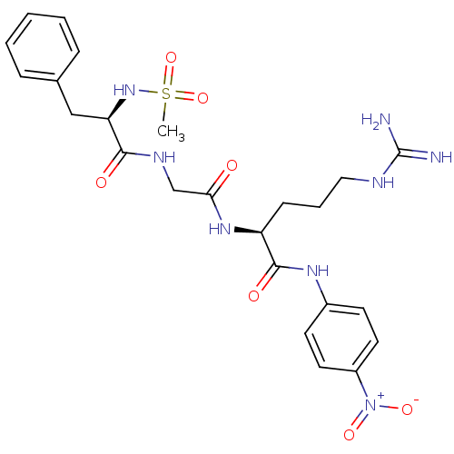 Chromozym tPA (2S)-5-carbamimidamido-2-{2-[(2R)-2-methanesulfonamido-3-phenylpropanamido]acetamido}-N-(4-nitrophenyl)pentanamide; acetic acid N-Methylsulfonyl-D-Phe-Gly-Arg-4-nitranilide acetate BDBM13553
 Tpa LPQTV BDBM20269 (2S)-N-[(1S,2S)-1-{[(1S)-1-carbamoyl-2-methylpropyl]carbamoyl}-2-hydroxypropyl]-2-{[(2S)-1-[(2S)-2-[(2S)-2-acetamido-3-[4-(2H-1,2,3,4-tetrazol-5-yl)phenyl]propanamido]-4-methylpentanoyl]pyrrolidin-2-yl]formamido}pentanediamide gp130 (904) derived peptide, 27
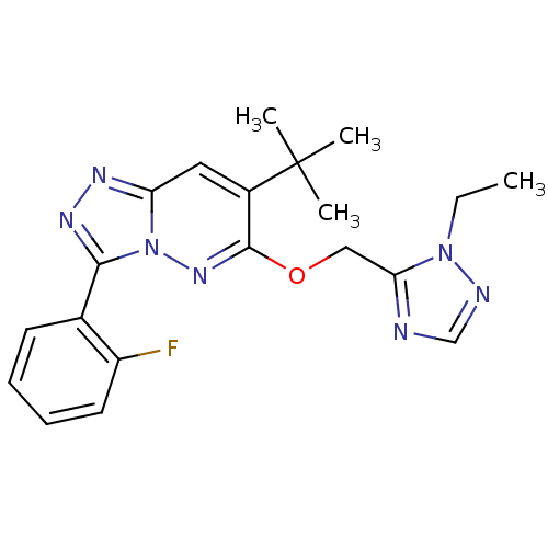 TPA-023 CHEMBL200177 7-(1,1-dimethyleth-1-yl)-3-(2-fluoro-phenyl)-6-(2-ethyl-2H-1,2,4-trizol-3-ylmethoxy)-[1,2,4]triazolo[4,3-b]pyridazine 7-tert-butyl-6-((2-ethyl-2H-1,2,4-triazol-3-yl)methoxy)-3-(2-fluorophenyl)-[1,2,4]triazolo[4,3-b]pyridazine BDBM50176771
- Qiu, C; Wei, R; Bian, J; Lin, X; Bai, T; He, J; Guo, X; Chu, Y Novel 4-triazole phenyl amide (4-TPA) molecules: Potent promoters of α-synuclein fibril disassembly. Eur J Med Chem 273:
- Renatus, M; Bode, W; Huber, R; Stürzebecher, J; Stubbs, MT Structural and functional analyses of benzamidine-based inhibitors in complex with trypsin: implications for the inhibition of factor Xa, tPA, and urokinase. J Med Chem 41: 5445-56 (1999)
- ChEBML_347237 Inhibition of tPA
- ChEBML_354130 Inhibition of tPA
- ChEBML_1769528 Inhibition of human TPA
- ChEBML_398432 Inhibition of recombinant tPA
- ChEMBL_450237 (CHEMBL900512) Inhibition of tPA
- ChEMBL_469233 (CHEMBL948934) Inhibition of tPA
- ChEMBL_552760 (CHEMBL954038) Inhibition of tPA
- ChEMBL_599100 (CHEMBL1047155) Inhibition of tPA
- ChEMBL_1461147 (CHEMBL3395472) Inhibition of human TPA
- ChEMBL_1623084 (CHEMBL3865436) Inhibition of human TPA
- ChEMBL_1795369 (CHEMBL4267486) Inhibition of human tPA
- ChEMBL_1795378 (CHEMBL4267495) Inhibition of mouse tPA
- ChEMBL_1898092 (CHEMBL4400127) Inhibition of human TPA
- ChEMBL_326973 (CHEMBL859774) Binding affinity to tPA
- ChEMBL_447832 (CHEMBL898082) Inhibition of human tPA
- ChEMBL_461717 (CHEMBL928848) Inhibition of human tPA
- ChEMBL_465215 (CHEMBL931927) Inhibition of human tPA
- ChEMBL_595930 (CHEMBL1041783) Inhibition of human tPA
- ChEMBL_937198 (CHEMBL2318524) Inhibition of human tPA
- ChEMBL_1463566 (CHEMBL3398784) Inhibition of TPA (unknown origin)
- ChEMBL_1499039 (CHEMBL3584636) Inhibition of tPA (unknown origin)
- ChEMBL_1671577 (CHEMBL4021606) Inhibition of TPA (unknown origin)
- ChEMBL_1921592 (CHEMBL4424437) Inhibition of tPA (unknown origin)
- ChEMBL_452507 (CHEMBL902746) Inhibition of human recombinant tPA
- ChEMBL_208240 (CHEMBL872720) Inhibitory activity against serine protease TPA
- ChEMBL_470233 (CHEMBL935743) Inhibition of human tPA after 30 mins
- ChEMBL_207472 (CHEMBL816753) Binding affinity was evaluated on serine protease TPA.
- ChEMBL_1458012 (CHEMBL3367186) Inhibition of TPA-induced AP-1 activation (unknown origin)
- ChEMBL_208241 (CHEMBL815719) Inhibitory activity against human serine protease (TPA) (2.5*10e6*)
- ChEMBL_423773 (CHEMBL853712) Inhibition of TPA-induced ODC activity in T24 cells
- ChEMBL_790755 (CHEMBL1926208) Inhibition of human TPA by Michaelis-Menten equation analysis
- ChEMBL_1290685 (CHEMBL3117488) Inhibition of transcription factor AP-1 binding to oligonucleotide containing TPA-responsive element in TPA-activated human HeLa cells after 1 hr by ELISA
- ChEMBL_423435 (CHEMBL909394) Inhibition of TPA-induced ornithine decarboxylase expressed in T24 cells
- ChEMBL_766825 (CHEMBL1827858) Inhibition of AP1 expressed in HEK293T cells coexpressing beta-lactamase pre-incubated 1 hr before TPA addition measured 18 hrs post TPA challenge by fluorimetric assay
- ChEMBL_1814631 (CHEMBL4314205) Inhibition of human tPA using S-2586 as spectrozyme tPA substrate incubated for 15 mins followed by substrate addition and measured after 1 hr by chromogenic assay
- ChEMBL_1661165 (CHEMBL4010777) Inhibition of TACE in NHEK assessed as reduction in LPS/TPA-stimulated TNFalpha production preincubated for 1 hr followed by LPS/TPA stimulation for 24 hrs by HTRF assay
- ChEMBL_1751369 (CHEMBL4186129) Inhibition of TACE in NHEK assessed as reduction in LPS/TPA-stimulated TNFalpha production preincubated for 1 hr followed by LPS/TPA stimulation for 24 hrs by HTRF assay
- ChEMBL_759677 (CHEMBL1811510) Inhibition of TPA-induced AP-1 activity expressed in human HEK293T cells treated 1 hr before TPA challenge measured after 18 hrs by FRET based beta-lactamase reporter assay
- ChEMBL_1923129 (CHEMBL4426085) Inhibition of human thrombin using pefachrom tPA as substrate by spectrophotometric method
- ChEMBL_1987858 (CHEMBL4621405) Inhibition of human tPa using fluorescent peptide as substrate by florescence assay
- ChEMBL_329857 (CHEMBL870579) Inhibition of the expression of AP1-luciferase by TPA-stimulated NIH3T3 cells
- ChEMBL_854020 (CHEMBL2155317) Inhibition of tPA assessed as S-2288 substrate hydrolysis by microplate reader analysis
- ChEMBL_573443 (CHEMBL1061338) Inhibition of TPA-induced AP1 transfected in HEK293 cells assessed as inhibition of beta-lactamase reporter activity treated 1 hr before TPA stimulation measured after 18 hrs by luciferase reporter gene assay
- ChEMBL_811901 (CHEMBL2013271) Inhibition of tPA using CH3SO2-D-HHT-Gly-Arg-pNA as substrate by spectrofluorometry
- ChEMBL_1580947 (CHEMBL3812615) Inhibition of human thrombin using pefachrom tPa as substrate after 3 mins by photometric method
- ChEMBL_310412 (CHEMBL834039) Repression of AP1-luciferase reporter activity in TPA-stimulated U2OS cells expressing rat glucocorticoid receptor
- ChEMBL_341910 (CHEMBL863833) Inhibition of cPLA2-alpha activity assessed by TPA-induced arachidonic acid release in human platelet
- ChEMBL_1856672 (CHEMBL4357401) Inhibition of activated human tPA using methylsulfonyl-D-cyclohexylalanyl-Gly-Arg-pNA as substrate by spectrophotometry
- ChEMBL_310419 (CHEMBL834045) Repression of NF-kappaB-luciferase reporter activity in TPA stimulated U2OS cells expressing rat glucocorticoid receptor
- ChEMBL_1540267 (CHEMBL3739098) Inhibition of mouse tPA after 15 mins using Z-D-Arg-Gly-Arg-p-nitroanilide as substrate
- ChEMBL_1925806 (CHEMBL4428878) Inhibition of tPA (unknown origin) incubated for 30 mins measured for 7 mins by morrison plot analysis
- ChEMBL_1458005 (CHEMBL3367179) Inhibition of AP-1 (unknown origin) expressed in TPA-stimulated mouse NIH3T3 cells by luciferase reporter gene assay
- ChEMBL_1933772 (CHEMBL4479424) Inhibition of human alpha-thrombin using chromogenic substrate Pefachrome tPa measured every second for 20 mins by spectrophotometry
- ChEMBL_1296325 (CHEMBL3129584) Binding affinity to human tPA assessed as release of p-nitroaniline after 10 to 120 mins by spectrophotometric analysis
- ChEMBL_475613 (CHEMBL923246) Inhibition of cPLA2-alpha in TPA-stimulated intact human platelets assessed as liberation of arachidonic acid after 60 mins
- ChEMBL_776474 (CHEMBL1913571) Stabilization of Pdcd4 expressed in human HEK293 cells assessed as inhibition of TPA-induced degradation by luciferase reporter assay
- ChEMBL_949240 (CHEMBL2345659) Inhibition of human tPA using Z-Gly-Gly-Arg-AMC as substrate assessed as residual activity by fluorometric analysis
- ChEMBL_1475712 (CHEMBL3424914) Inhibition of human alpha-thrombin using tPa (CH3SO2-D-Cha-Gly-Arg-pNA.AcOH) as chromogenic substrate by kinetic photometric assay
- ChEMBL_1871027 (CHEMBL4372194) Inhibition of tPA (unknown origin) using Z-Pyr-Gly-Arg-MCA as substrate after 30 mins relative to untreated control
- Fluorometric PAI-1/tPA IC50 Plate Assay at pH 7.8 PAI-1 inhibitor compounds were dissolved in DMSO to a final concentration of (10-50 mM), depending upon solubility. Compounds were then diluted in physiologic buffer (40 mM HEPES, 100 mM NaCl, 0.05% Tween-20, pH7.8) containing (10% DMSO and a dilution series (from 0 to 1000 μM depending on solubility) was prepared. 80 μL of compound was added per well to a 96-well black, opaque microplate in duplicate. 10 μL of 20 nM recombinant active human PAI-1 (Molecular Innovations) in physiologic buffer, as set out above, was added per well and the mixture was agitated for 15 minutes at room temperature. 10 uL of 25 nM human tissue type PA (tPA) (Activase (alteplase), Genentech) was added per well and the plate was agitated for an additional 30 minutes at room temperature. Tissue type PA activity in each reaction mixture was determined by adding Phe-Gly-Arg-AMC fluorogenic substrate (100 μL of 100 μM) (Centerchem). The rate of AMC release by tPA was measured at an excitation wavelength of 370 nm and an emission wavelength of 440 nm. Controls included PAI-1 and tPA in the absence of compound and tPA alone. Percent PAI-1 inhibition was calculated using the following formula: [(tPA alone-tPA/PAI-1+ compound)/(tPA alone-PAI-1/tPA)]*100%. The IC50 is calculated using Graphit (IC50 0-100%).
- ChEBML_1564078 Inhibition of human TPA assessed as reduction in release of p-nitroaniline after 10 to 120 mins by Michaelis-Menten equation analysis
- ChEMBL_1363410 (CHEMBL3295545) Binding affinity to full length Glu-plasminogen in human plasma assessed as inhibition of interaction with fibrin in presence of tPA
- ChEMBL_1765365 (CHEMBL4200612) Inhibition of tPA (unknown origin) using chromogenic H-D-Ile-L-Pro-L-Arg-p-nitoraniline as substrate by fluorescence assay
- ChEMBL_1564078 (CHEMBL3783219) Inhibition of human TPA assessed as reduction in release of p-nitroaniline after 10 to 120 mins by Michaelis-Menten equation analysis
- ChEMBL_1634335 (CHEMBL3877127) Inhibition of tPA (unknown origin) using Methylsulfonyl-D-Phe-Gly-Arg-pNA as substrate preincubated for 30 mins followed by substrate addition
- ChEMBL_1692237 (CHEMBL4042886) Transrepression activity at glucocorticoid receptorin human HeLa cells assessed as inhibition of TPA-induced collagenase promoter activity by luciferase reporter gene assay
- ChEBML_1681819 Inhibition of human TPA using methylsulfonyl-D-cyclohexylalanyl-Gly-Arg-pNA as substrate at 37 degC after 10 to 120 mins by spectrophotometric method
- ChEMBL_1543047 (CHEMBL3743970) Inhibition of recombinant human tPA using H-D-Ile-Pro-L-Arg-pNA-2HCl as substrate measured for 5 mins by spectrophotometric assay
- ChEMBL_655624 (CHEMBL1244668) Inhibition of Myc-tagged human Dusp6 expressed in TPA-induced human HeLa cells assessed as decrease in dephosphorylation of pERK measured by immunostaining
- ChEMBL_655625 (CHEMBL1244669) Inhibition of Myc-tagged human Dusp1 expressed in TPA-induced human HeLa cells assessed as decrease in dephosphorylation of pERK measured by immunostaining
- ChEMBL_508044 (CHEMBL945487) Inhibition of LFA1/ICAM1-mediated CFSE-labeled TPA-stimulated human HL60 cell adhesion to ICAM1 expressing human HeLa cells after 45 mins by fluorescence analysis
- ChEMBL_741821 (CHEMBL1769966) Inhibition of TPA-induced AP-1 activation in cells expressing beta-lactamase after 18 hrs using FRET substrate by cell-based high-throughput screening assay
- ChEMBL_1612873 (CHEMBL3854673) Inhibition of human recombinant tPA using methylsulfonyl-D-cyclohexylalanylGly-Arg-pNA as substrate assessed as release of pNA after 10 to 120 mins by spectrophotometric method
- ChEMBL_2134852 (CHEMBL4844462) Inhibition of recombinant human tPA using H-D-Isoleucyl-L-prolyl-L-arginine p-nitroaniline dihydrochloride as substrate measured after 5 mins by plate reader analysis
- ChEMBL_2426277 Inhibition of tPA (unknown origin) using H-D-lle-Pro-Arg-pNA-2HCl as chromogenic substrate incubated for 20 mins followed by substrate addition by spectrophotometric analysis
- ChEMBL_664245 (CHEMBL1261752) Transrepression activity at glucocorticoid receptor-mediated antiinflammatory activity in human HeLa cells assessed as inhibition of TPA-induced collagenase promoter activity by luciferase reporter gene assay
- ChEMBL_801832 (CHEMBL1947444) Inhibition of recombinant tPA using H-D-Ile-Pro-Arg-pNA.2HCl as substrate preincubated for 15 mins prior substrate addition measured for 10 mins by spectrophotometry
- ChEMBL_555519 (CHEMBL963779) Inhibition of glucocorticoid receptor-glucocorticoid complex transfected in rat R1 cells assessed as down-regulation of TPA-responsive element proinflammatory gene expression after 24 hrs by beta-galactosidase assay
- ChEMBL_63593 (CHEMBL675240) Inhibition of the heparin binding epidermal growth factor (HB-EGF) release from fibrosarcoma HT1080 transfectants expressing alkaline phosphate (AP) tagged HB-EGF stimulated by 12-O-tetradecanoylphorbol 13-acetate(TPA).
- ChEMBL_555521 (CHEMBL963781) Inhibition of glucocorticoid receptor-glucocorticoid complex transfected in rat R1 cells assessed as down-regulation of TPA-responsive element proinflammatory gene expression at 10 uM after 24 hrs by beta-galactosidase assay
- ChEMBL_751484 (CHEMBL1787264) Inhibition of TPA-induced degradation of Pdcd4 (amino acid 39-91) expressed in human HEK293 cells assessed as minimum compound concentration required for 50% recovery of Pdcd4-luciferase signal incubated for 8 hrs by luciferase reporter gene assay
- ChEMBL_1363409 (CHEMBL3295544) Binding affinity to human full length Glu-plasminogen in buffer assessed as inhibition of interaction with fibrin preincubated for 15 mins followed by thrombin addition measured every 2 mins for 15 hrs by Spectra-Max reader analysis in presence of tPA
- ChEMBL_1752854 (CHEMBL4187614) Inhibition of human PAI-1 assessed as remaining enzyme activity by measuring p-nitroaniline release pre-incubated for 15 mins before 9.8 nM human tPA substrate addition for 15 mins using 67 nM enzyme and chromogenic substrate S-2288 by spectrophotometry
- ChEMBL_1752858 (CHEMBL4187618) Inhibition of human PAI-1 assessed as remaining enzyme activity by measuring p-nitroaniline release pre-incubated for 15 mins before 2 nM Spectrozyme tPA substrate addition for 15 mins using 5 nM enzyme and chromogenic substrate S-2288 by spectrophotometry
- ChEMBL_1752859 (CHEMBL4187619) Inhibition of human PAI-1 assessed as remaining enzyme activity by measuring p-nitroaniline release pre-incubated for 15 mins before 9.8 nM human tPA substrate addition for 15 mins using 24.5 nM enzyme and chromogenic substrate S-2288 by spectrophotometry
- Inhibition Assay Inhibition of human FIIa was determined by the method described in [0092]-[0098] using human alpha-thrombin from Enzyme Research Laboratories at 0.1 NIH U/mL and Mes-d-Cha-Gly-Arg-pNA (Pefachrome tPA) at 2 mM, 1 mM, and 0.5 mM as substrate; results are reported as Ki values (nanomolar).
- Inhibition Assay Inhibition of human t-PA was determined by the method described in [0092]-[0098] using recombinant human tissue-type plasminogen activator (Actilyse®) from Boehringer Ingelheim at 290 U/mL and Mes-d-Cha-Gly-Arg-pNA (Pefachrome tPA) at 4 mM, 2 mM, and 1 mM as substrate; results are reported as Ki values (nanomolar).
- Enzymatic Activity Assay Determination of the Selectivity To demonstrate the selectivity of the substances with respect to thrombin and factor Xa inhibition, the test substances are examined for their inhibition of other human serine proteases, such as factor factor XIa, trypsin, plasmin, tissue plasminogen activator (TPA), and plasma kallikrein. The determinations are carried out in microtitre plates. To determine the enzymatic activity of factor XIa (0.15 nmol/l from Kordia), trypsin (42 mU/ml from Sigma), plasmin (0.1 μg/ml from Kordia), TPA (1 nmol/l from Kordia) and plasma kallikrein (0.2 nmol/l from Loxo), these enzymes are dissolved (50 mmol/l of Tris buffer [C,C,C-tris(hydroxymethyl)aminomethane], 100 mmol/l of sodium chloride, 0.1% BSA [bovine serum albumin], 5 mmol/l of calcium chloride, pH 7.4) and incubated for 15 min with test substance in various concentrations in dimethyl sulphoxide and also with dimethyl sulphoxide without test substance. The enzymatic reaction is then started by addition of the appropriate substrates (5 μmol/l of Boc-Glu(OBzl)-Ala-Arg-AMC from Bachem for factor XIa, 5 μmol/l of Boc-Ile-Glu-Gly-Arg-AMC from Bachem for Trypsin, 50 μmol/l of MeOSuc-Ala-Phe-Lys-AMC from Bachem for plasmin, 5 μmol/l of CH3SO2-D-Phe-Gly-Arg-AMC from Pentapharm for TPA and 5 μmol/l of H-Pro-Phe-Arg-AMC from Bachem for plasma kallikrein). After an incubation time of 30 min at 22° C., fluorescence is measured (excitation: 360 nm, emission: 460 nm).
- Enzyme Inhibition Assay Kinetic inhibition of human thrombin was determined photometrically at 405 nm using the chromogenic substrate Pefachrom tPa. Reactions were performed under the conditions using different concentrations of substrate (182, 91 and 45 uM) and inhibitor (36.4, 27.3, 18.2 and 9.1 uM for the weakest inhibitor and 3.6, 2.7, 1.8 and 0.9 nM for the tightest binder). Activity of thrombin was adjusted by diluting solution with 154 mM NaCl until linear conversion of the substrate could be detected over 5 min in an appropriate absorption window (~0.2-0.8). The assay was stopped after 3 min with concentrated acetic acid and absorption in each well was corrected for the blank value. Ki values were determined as described by Dixon.
- Biacore Assay The test compounds were dissolved in 1% acetic acid and further complemented with an equal volume of DMSO. The resulting stock solutions were serially diluted in 0.5% acetic acid/50% DMSO. 1 μL aliquots of these solutions were placed into 384 well microplates (Greiner, black, transparent bottom), followed by 30 μL of diluted human citrated plasma (platelet-poor, final concentration: 5%; supplemented with fibrinogen, final concentration: 3 μM; dilution buffer: 20 mM HEPES, 150 mM NaCl, 0.01% Brij (pH 7)). The reactions were started by addition of 20 L of CaCl2 (final concentration: 10 mM), and tPA (tissue plasminogen activator, final concentration: 0.2 nM) in dilution buffer, followed by an additional volume of 20 μL dilution buffer for improved mixing. The reactions were incubated at 37° C. Clot formation and degradation was monitored spectrophotometrically by kinetic optical density measurements at 405 nm.
- Plasma-Based Clot Lysis Assay The clot-lysis test system configures the kinetics of clot formation and degradation in vitro and allows quantifying modulation of the process by selected test compounds.The test compounds were dissolved in 1% acetic acid and further complemented with an equal volume of DMSO. The resulting stock solutions were serially diluted in 0.5% acetic acid/50% DMSO. 1 μL aliquots of these solutions were placed into 384 well microplates (Greiner, black, transparent bottom), followed by 30 μL of diluted human citrated plasma (platelet-poor, final concentration: 5%; supplemented with fibrinogen, final concentration: 3 μM; dilution buffer: 20 mM HEPES, 150 mM NaCl, 0.01% Brij (pH 7)). The reactions were started by addition of 20 μL of CaCl2 (final concentration: 10 mM), and tPA (tissue plasminogen activator, final concentration: 0.2 nM) in dilution buffer, followed by an additional volume of 20 μL dilution buffer for improved mixing. The reactions were incubated at 37° C. Clot formation and degradation was monitored spectrophotometrically by kinetic optical density measurements at 405 nm. IC50 values were determined by comparing the resulting time courses with the time course of a blank control reaction.
- Plasma-based Clot Lysis Assay The clot-lysis test system configures the kinetics of clot formation and degradation in vitro and allows quantifying modulation of the process by selected test compounds.The test compounds were dissolved in 1% acetic acid and further complemented with an equal volume of DMSO. The resulting stock solutions were serially diluted in 0.5% acetic acid/50% DMSO. 1 μL aliquots of these solutions were placed into 384 well microplates (Greiner, black, transparent bottom), followed by 30 μL of diluted human citrated plasma (platelet-poor, final concentration: 5%; supplemented with fibrinogen, final concentration: 3 μM; dilution buffer: 20 mM HEPES, 150 mM NaCl, 0.01% Brij (pH 7)). The reactions were started by addition of 20 μL of CaCl2 (final concentration: 10 mM), and tPA (tissue plasminogen activator, final concentration: 0.2 nM) in dilution buffer, followed by an additional volume of 20 μL dilution buffer for improved mixing. The reactions were incubated at 37° C. Clot formation and degradation was monitored spectrophotometrically by kinetic optical density measurements at 405 nm. IC50 values were determined by comparing the resulting time courses with the time course of a blank control reaction.