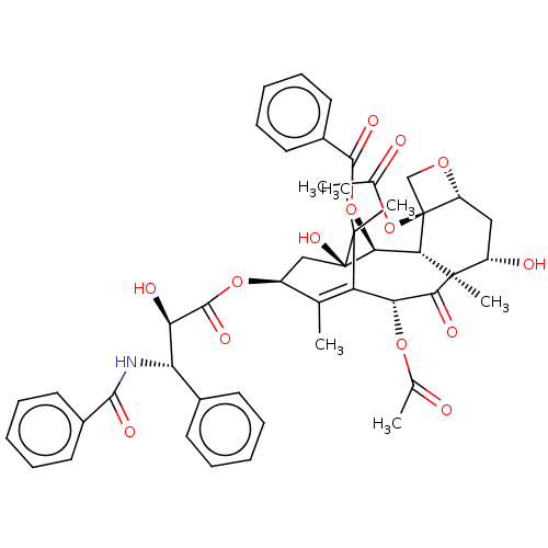
 taxol CHEMBL428647 BDBM50001839 PACLITAXEL
- Kwiatkowski, N; Deng, X; Wang, J; Tan, L; Villa, F; Santaguida, S; Huang, HC; Mitchison, T; Musacchio, A; Gray, N Selective aurora kinase inhibitors identified using a taxol-induced checkpoint sensitivity screen. ACS Chem Biol 7: 185-96 (2012)
- ChEMBL_837054 (CHEMBL2076139) TP_TRANSPORTER: inhibition of Taxol transepithelial transport (basal to apical) in Caco-2 cells
- ChEMBL_838522 (CHEMBL2078150) TP_TRANSPORTER: inhibition of Taxol transepithelial transport (basal to apical) in Caco-2 cells
- ChEMBL_1361313 (CHEMBL3293265) Inhibition of p-glycoprotein in human MDA435/LCC6MDR cells assessed as concentration required to reduce taxol IC50 by half after 5 days by MTS assay
- ChEMBL_2571444 Inhibition of MPS1 in human taxol-arrested U2OS cells assessed as effect on histone H3 phosphorylation at ser10 residue incubated for 2 hrs by arrayscan analysis
- CYP450 Inhibition Assay The ability of the R and S enantiomers of (4-fluorophenyl)(4-((5-methyl-1H-pyrazol-3-yl)amino)quinazolin-2-yl)methanol to inhibit the common drug metabolizing isoforms of cytochrome P450 (CYP) was evaluated against the following isoforms: CYP1A2, CYP2B6, CYP2C8, CYP2C9, CYP2C19, CYP2D6, and CYP3A4. The compounds were incubated in duplicate with eight test compound concentrations (final DMSO concentration of 0.20%) with human liver microsomes (0.25 or 0.50 mg/mL) and NADPH (1 mM) in the presence of CYP isoform specific probe substrates (phenacetin, bupropion, taxol, diclofenac, mephenyloin, dextromethorphan, testosterone) at the Km for 10-20 minutes at 37° C. Selective CYP isoform inhibitors (furafulline, ticlopidine, quercetin, sulfaphenazole, ticlopidine, quinidine, ketoconazole) were screened alongside the test compounds as positive controls.
- Binding Kinetics to KIF18a-Microtubule Complex Table 10: Compound binding kinetics parameters (kon and koff) were determined by the method of global progress curve analysis (GPCA). KIF18A (0.25 nM) was incubated for up to 24 hr with serially diluted compound in the assay buffer containing 80 mM PIPES, pH 6.9, 1 mM ATP, 0.1 mg/ml preformed microtubule from porcine brain (Cytoskeleton), 1 mM MgCl2, 1 μM Taxol, 75 mM KCl, 1 mM EGTA, 1 mM DTT, 0.01% BSA and 0.005% Tween-20. ADP product levels were determined by the Promega ADP-Glo assay. The time/dose-dependent progress curves were then globally fit to a Michaelis-Menten kinetics model with 1-step slow binding inhibition to derive both on-rate kon and off-rate koff values.
- Binding Kinetic Assay Compound binding kinetics parameters (kon and koff) were determined by the method of global progress curve analysis (GPCA). KIF18A (0.25 nM) was incubated for up to 24 hr with serially diluted compound in the assay buffer containing 80 mM PIPES, pH 6.9, 1 mM ATP, 0.1 mg/ml preformed microtubule from porcine brain (Cytoskeleton), 1 mM MgCl2, 1 M Taxol, 75 mM KCl, 1 mM EGTA, 1 mM DTT, 0.01% BSA and 0.005% Tween-20. ADP product levels were determined by the Promega ADP-Glo assay. The time/dose-dependent progress curves were then globally fit to a Michaelis-Menten kinetics model with 1-step slow binding inhibition to derive both on-rate kon and off-rate koff values (Zhang, R., Wong, K. (2017): High performance enzyme kinetics of turnover, activation and inhibition for translational drug discovery , Expert Opinion on Drug Discovery, 2017 January; 12(1):17-37. doi: 10.1080/17460441.2017.1245721).
- KIF18A Biochemical Assay A KIF18A ATPase assay was performed in small-volume, nonbinding, 384-well white plates at a final volume of 10 μL/well. Test compounds (10 mM solution in DMSO; 100 nL/well) were serially diluted 3-fold over 10-point concentration range. A solution of KIF18A (0.4 nM, 5 μL/well; 1-367) in assay buffer (15 mM Tris-HCl [pH 7.5](Boston Bioproducts Inc), 10 mM MgCl2 (Boston Bioproducts Inc), 0.01% Pluronic F-68 (Gibco Inc), 1 uM Taxol (Cytoskeleton Inc), 30 mg/ml pre-formed porcine Microtubules (Cytoskeleton Inc)). The reaction was initiated by the addition of 5 μL of substrate solution (10 μM Ultra-Pure ATP in assay buffer) into the wells. The plates were incubated at room temperature for 45 minutes. After the indicated incubation times, 10 μL ADP-Glo reagent was added to the reactions and the plate was incubated at room temperature for 40 min. Then, 20 μL of kinase detection reagent was added and after an incubation time of 40 min, luminescence was recorded on Envision plate reader (Perkin Elmer, Billerica, MA).
- Inhibition Assay The motor domain of the human kinesin spindle protein KSP/Eg5 (tebu-bio/Cytoskeleton Inc, No. 027EG01-XL) was incubated in a concentration of 10 nM with microtubuli (bovine or porcine, tebu-bio/Cytoskeleton Inc) stabilized with 50 μg/ml taxol (Sigma No. T7191-5MG) for 5 min at RT in 15 mM PIPES, pH 6.8 (5 mM MgCl2 and 10 mM DTT, Sigma). The freshly prepared mixture was aliquoted into a 384 MTP (Greiner bio-one REF 781096). The inhibitors to be examined at concentrations of 1.0×10⁻⁶ M to 1.0×10⁻¹³ M and ATP (final concentration 500 μM, Sigma) were then added. Incubation was at RT for 2 h. ATPase activity was detected by detecting the inorganic phosphate formed using malachite green (Biomol). After addition of the reagent, the assay was incubated at RT for 50 min prior to detection of the absorption at a wavelength of 620 nm. The positive controls used were monastrol (Sigma, M8515-1 mg) and ispinesib (AdooQ Bioscience A10486). The individual data of the dose-activity curve are eight-fold determinations.
- Assay for Inhibition of KIF11 The ATPase rate for Kif11 was monitored using an enzyme-coupled assay. Porcine brain microtubules (Cat #MT002) were purchased from Cytoskeleton (Denver, CO) and polymerized as per manufacturer's instructions and stored at 1 mg/ml at −80° C. GST-tagged Kif11/Eg5 (Cat #EG01-XL) and the Kinesin ELIPA Biochem Kit (Cat #BK060) was also purchased from Cytoskeleton. Taxol, microtubules, 7-methylthioguanosine (MESG), and purine nucleoside phosphorylase (PNP) were added to final concentrations of 15 mM, 0.05 mg/mL, 200 mM, 1U/m, 1 mM in EPLIA Reaction Buffer and allowed to incubate at room temperature for 15 minutes under rocking. Kif11 was then added at a final concentration of 0.025 mM and allowed to incubate at room temperature for an additional 15 minutes. Master mix (18.5 mL) was added to a black 384-well plate (Corning 3575), to which 0.5 mL of DMSO or compound were added to appropriate wells. Plates were incubated at room temperature for 2 hours. ATPase reactions were initiated by addition of 1 mM ATP and monitored by OD340 every 1 minute over the course of 2 hours. The rate of the reaction was taken from the linear part of the resulting curve. IC50 was determined by plotting inhibitor concentration v. ATPase rate.
- Inhibition of KIF18A Microtubule-Dependent ATPase Activity Table 9: Test compounds were plated in a 3× dilution scheme in a 384-well plate. Assay buffer: 80 mM PIPES (pH 6.9), 1 mM MgCl2, 75 mM KCl, 1 mM EGTA, 1 mM DTT, 0.01% BSA, 0.005% Tween-20, 1 μM Taxol in H2O. To 50 nL of compound in DMSO was added 2.5 μL of enzyme mix [4 nM hKIF18A (1-374) in assay buffer]. After incubation at room temperature for 30 min, 2.5 μL of microtubule mix was added [0.2 mg/mL pre-formed microtubules, 2.0 mM ATP in assay buffer], the plate was centrifuged for 30 s and then incubated at 28° C. for 60 min. 5 μL of Promega ADP-Glo Max R1 was added, the plate was centrifuged for 30 s, and the mixture incubated for 4 h at room temperature. 10 μL of Promega ADP-Glo Max R2 was added, the plate centrifuged for 30 s, and incubated for 60 min at room temperature. Luminescence was measured with an Envision plate reader, and % Inhibition was calculated for each well as: ([max−min]−[test−min])/[max−min]. IC50 values were calculated from concentration vs. % Inhibition data via a four-parameter variable slope model.
- Inhibition of KIF18A Microtubule-Dependent ATPase Activity Test compounds were plated in a 3x dilution scheme in a 384-well plate. Assay buffer: 80 mM PIPES (pH 6.9), 1 mM MgCl2, 75 mM KCl, 1 mM EGTA, 1 mM DTT, 0.01% BSA, 0.005% Tween-20, 1 uM Taxol in H2O. To 50 nL of compound in DMSO was added 2.5 uL of enzyme mix [4 nM hKIF18A (1-374) in assay buffer]. After incubation at room temperature for 30 min, 2.5 uL of microtubule mix was added [0.2 mg/mL pre-formed microtubules, 2.0 mM ATP in assay buffer], the plate was centrifuged for 30 s and then incubated at 28 C. for 60 min. 5 uL of Promega ADP-Glo Max R1 was added, the plate was centrifuged for 30 s, and the mixture incubated for 4 h at room temperature. 10 uL of Promega ADP-Glo Max R2 was added, the plate centrifuged for 30 s, and incubated for 60 min at room temperature. Luminescence was measured with an Envision plate reader, and % Inhibition was calculated for each well as: ([max-min]-[test-min])/[max-min]. IC50 values were calculated from concentration vs. % Inhibition data via a four-parameter variable slope model.
- Inhibition of the Kinesin Spindle Protein KSP/Eg5 The motor domain of the human kinesin spindle protein KSP/Eg5 (from tebu-bio/Cytoskeleton Inc, No. 027EG01-XL) is incubated at a concentration of 10 nM with 50 μg/ml taxol- (from Sigma No. T7191-5MG) stabilized microtubuli (bovine or porcine, from tebu-bio/Cytoskeleton Inc) for 5 min at RT in 15 mM PIPES, pH 6.8 (5 mM MgCl2 and 10 mM DTT, from Sigma). The freshly prepared mixture was aliquoted into a 384-well MTP. The inhibitors to be examined at concentrations of 1.0×10⁻⁶ M to 1.0×10⁻¹³ M and ATP (final concentration 500 μM, from Sigma) were then added. Incubation was carried out at RT for 2 h. ATPase activity was detected by detecting the inorganic phosphate formed using malachite green (from Biomol). After addition of the reagent, the assay was incubated at RT for 50 minutes prior to detection of the absorption at a wavelength of 620 nm. Monastrol (Fa. Sigma, M8515-1 mg) and Ispinesib (from Adooq A10486) were used as positive control. The individual data of the dose-activity curve are octuple determinations. The IC50 values are means of three independent experiments. The 100% control was the sample which had not been treated with inhibitors.
- Inhibition of the Kinesin Spindle Protein KSP/Eg5 The motor domain of the human kinesin spindle protein KSP/Eg5 (tebu-bio/Cytoskeleton Inc, No. 027EG01-XL) was incubated in a concentration of 10 nM with microtubuli (bovine or porcine, tebu-bio/Cytoskeleton Inc) stabilized with 50 μg/ml taxol (Sigma No. T7191-5MG) for 5 min at RT in 15 mM PIPES, pH 6.8 (5 mM MgCl2 and 10 mM DTT, Sigma). The freshly prepared mixture was aliquoted into a 384 MTP (from Corning). The inhibitors to be examined at concentrations of 1.0×10⁻⁶ M to 1.0×10⁻¹³ M and ATP (final concentration 500 μM, Sigma) were then added. Incubation was at RT for 2 h. ATPase activity was detected by detecting the inorganic phosphate formed using malachite green (Biomol). After addition of the reagent, the assay was incubated at RT for 50 min prior to detection of the absorption at a wavelength of 620 nm. The positive controls used were monastrol (Sigma, M8515-1 mg) and ispinesib (AdooQ Bioscience A10486). The individual data of the dose-activity curve are eight-fold determinations. The IC50 values are means of two independent experiments. The 100% control was the sample which had not been treated with inhibitors.
- Enzyme Assay Microtubule-stimulated ATPase activity assay is used to measure KIF18A enzyme activity after treatment with compound. Compounds were 2-fold serially diluted in DMSO (Sigma Inc) over 22-point concentration range. Recombinant human KIF18A (1-467 His-tagged) protein was expressed using a baculovirus system and purified by affinity chromatography by Amgen Inc. Concentrations of KIF18A protein, microtubules (MT), and ATP in the reaction were optimized for standardized homogenous enzyme assay using ADP-Glo Kinase/ATPase Assay Kit (Promega Inc). The assay measures ADP formed from the ATPase reaction. Prepare reaction buffer [(15 mM Tris, pH 7.5 (Teknova Inc), 10 mM MgCl2 (JT Baker Inc), 0.01% Pluronic F-68 (Life Technologies Inc), 1 μM Taxol (Cytoskeleton Inc), and 30 μg/mL pig microtubules (Cytoskeleton Inc)]. Add compound and KIF18A protein (30 nM) to prepared reaction buffer and incubated for 15 minutes at room temperature, next add ATP (at Km, 75 μM) to the reaction mixture and incubated for an additional 15 minutes at room temperature. Mix 5 μl of ADP-Glo™ Reagent and 2.5 μl of the reaction mixture and incubate for 40 minutes at room temperature. Add 10 μl ADP-Glo Detection Reagent and incubate for 40 minutes at room temperature. Read luminescence using EnVision microplate reader with ultra-luminescence module (Perkin Elmer Inc). Concentration-response curve-fitting and IC50 determination was performed using Genedata Screener Software (Standard 15.0.1, Genedata Inc) with a four-parameter logistic regression fit model.