 US10793575, Example ibrutinib BDBM50357312 US10835536, Ref. Ex. Comp 1 US20240358707, Compound 13 US11407754, Example Ibrutinib IBRUTINIB PCI-32765 US10124003, Ref. Ex. Compound 1 US20230364079, Example Ibrutinib US11186578, Example Ibrutinib US10711006, Compound Ibrutinib US10919899, Ibrutinib US9278100, 1 US9108973, Ref 1 US20240059694, Compound Ibrutinib US9181263, 1 US20240139326, Compound Ibrutinib US11078206, Example Ibrutinib US11339167, Example Ibrutinib
- Guo, M; Dai, S; Wu, D; Duan, Y; Li, J; Qu, L; Jiang, L; Chen, Z; Chen, X; Chen, Y Characterization of ibrutinib as a non-covalent inhibitor of SRC-family kinases. Bioorg Med Chem Lett 34: (2021)
- Ran, F; Liu, Y; Wang, C; Xu, Z; Zhang, Y; Liu, Y; Zhao, G; Ling, Y Review of the development of BTK inhibitors in overcoming the clinical limitations of ibrutinib. Eur J Med Chem 229: (2022)
- Long, K; Close, DA; Johnston, PA; Huryn, DM Replacement of the hydroxamic acid group in the selective HDAC8 inhibitor PCI-34051. Bioorg Med Chem Lett 108:
- Balasubramanian, S; Ramos, J; Luo, W; Sirisawad, M; Verner, E; Buggy, JJ A novel histone deacetylase 8 (HDAC8)-specific inhibitor PCI-34051 induces apoptosis in T-cell lymphomas. Leukemia 22: 1026-34
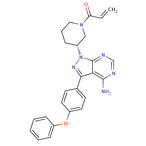
- In Vitro Inhibitory Activities on ITK In vitro activity in inhibiting ITK kinase was determined similar to that in respect of BTK kinase.The inhibitory activity of the compound of Example 9 on BTK is very high with an IC50 value of 1.9 nM, comparable to the literature compound, Ibrutinib/PCI-32765, which has been approved for clinical application. The platform for the enzymatic assay on ITK kinase (another Tec Kinase, mainly expressed in T cells) was establish with the same method, and the inhibitory ability of the compound of example 9 on ITK was tested. The results showed that IC50 value of this inhibition of ITK was more than 1000 nM. The selectivity of the compound of example 9 on BTK vs ITK was calculated as more than 1000 folds, while the selectivity of the literature compound, PCI-32765, was reported as approximately 100 folds. Accordingly, the selectivity of the compound of example 9 is significantly higher than that of PCI-32765.
- ChEMBL_41044 (CHEMBL883285) Compound was tested for inhibition of Beta-lactamase from Staphylococcus aureus PCI
- ChEMBL_2227009 (CHEMBL5140522) Inhibition of PDCD4 in human HeLa cells tranfected with pCI-neo-luciferase-PDCD4 3'-UTR plasmid assessed as increase in luciferase activity by luciferase reporter assay
- Kinetic Labeling Experiments of Ibrutinib Derivatives with BTK BTK kinase domain was expressed and purified as previously reported65. Binding experiments were performed in Tris 20 mM pH=8, 50 mM NaCl, and 1 mM DTT. BTK kinase domain was diluted to 2 μM in the buffer, and 2 μM Ibrutinib derivatives were added by adding 1/100th volume from a 200 μM solution. The reaction mixtures, at room temperature for various times, were injected into the LC/MS. For data analysis, the raw spectra were deconvoluted using a 20000:40000 Da window and 1 Da resolution. The signal from masses 20000:30000 and 33000:40000 (which contained no peaks) was averaged and subtracted from the whole signal. The peaks of each species were integrated using a 100 Da window in every direction (reducing the window down to 10 Da did not change the results significantly).
- Inhibition Assay A PDE10A assay may for example, be performed as follows: The assay is performed in 60 uL samples containing a fixed amount of the relevant PDE enzyme (sufficient to convert 20-25% of the cyclic nucleotide substrate), a buffer (50 mM HEPES7.6; 10 mM MgCl2; 0.02% Tween20), 0.1 mg/ml BSA, 225 pCi of 3H-labelled cyclic nucleotide substrate, tritium labeled cAMP to a final concentration of 5 nM and varying amounts of inhibitors. Reactions are initiated by addition of the cyclic nucleotide substrate, and reactions are allowed to proceed for one hr at room temperature before being terminated through mixing with 15 uL 8 mg/mL yttrium silicate SPA beads (Amersham). The beads are allowed to settle for one hr in the dark before the plates are counted in a Wallac 1450 Microbeta counter.
- Inhibition Assay A PDE10A assay may for example, be performed as follows: The assay is performed in 60 uL samples containing a fixed amount of the relevant PDE enzyme (sufficient to convert 20-25% of the cyclic nucleotide substrate); a buffer (50 mM HEPES7.6; 10 mM MgCl2; 0.02% Tween20), 0.1 mg/ml BSA, 225 pCi of 3H-labelled cyclic nucleotide substrate, tritium labeled cAMP to a final concentration of 5 nM and varying amounts of inhibitors. Reactions are initiated by addition of the cyclic nucleotide substrate, and reactions are allowed to proceed for one hr at room temperature before being terminated through mixing with 15 uL 8 mg/mL yttrium silicate SPA beads (Amersham). The beads are allowed to settle for one hr in the dark before the plates are counted in a Wallac 1450 Microbeta counter.
- Inhibition Assay A PDE9 assay may for example, be performed as follows: The assay is performed in 60 uL samples containing a fixed amount of the relevant PDE enzyme (sufficient to convert 20-25% of the cyclic nucleotide substrate), a buffer (50 mM HEPES 7.6; 10 mM MgCl2; 0.02% Tween20), 0.1 mg/ml BSA, 225 pCi of 3H-labelled cyclic nucleotide substrate, tritium labeled cAMP to a final concentration of 5 nM and varying amounts of inhibitors. Reactions are initiated by addition of the cyclic nucleotide substrate, and reactions are allowed to proceed for one hr at room temperature before being terminated through mixing with 15 uL 8 mg/mL yttrium silicate SPA beads (Amersham). The beads are allowed to settle for one hr in the dark before the plates are counted in a Wallac 1450 Microbeta counter.
- Inhibition Assay A PDE10A assay may for example, be performed as follows: The assay is performed in 60 uL samples containing a fixed amount of the relevant PDE enzyme (sufficient to convert 20-25% of the cyclic nucleotide substrate), a buffer (50 mM HEPES7.6; 10 mM MgCl2; 0.02% Tween20), 0.1 mg/ml BSA, 225 pCi of 3H-labelled cyclic nucleotide substrate, tritium labeled cAMP to a final concentration of 5 nM and varying amounts of inhibitors. Reactions are initiated by addition of the cyclic nucleotide substrate, and reactions are allowed to proceed for one hr at room temperature before being terminated through mixing with 15 uL 8 mg/mL yttrium silicate SPA beads (Amersham). The beads are allowed to settle for one hr in the dark before the plates are counted in a Wallac 1450 Microbeta counter. The measured signal can be converted to activity relative to an uninhibited control (100%) and IC50 values can be calculated using the Xlfit extension to EXCEL.
- Inhibition Assay A PDE10A assay may for example, be performed as follows: The assay is performed in 60 uL samples containing a fixed amount of the relevant PDE enzyme (sufficient to convert 20-25% of the cyclic nucleotide substrate), a buffer (50 mM HEPES7.6; 10 mM MgCl2; 0.02% Tween20), 0.1 mg/ml BSA, 225 pCi of 3H-labelled cyclic nucleotide substrate, tritium labeled cAMP to a final concentration of 5 nM and varying amounts of inhibitors. Reactions are initiated by addition of the cyclic nucleotide substrate, and reactions are allowed to proceed for one hr at room temperature before being terminated through mixing with 15 uL 8 ng/mL yttrium silicate SPA beads (Amersham). The beads are allowed to settle for one hr in the dark before the plates are counted in a Wallac 1450 Microbeta counter. The measured signal can be converted to activity relative to an uninhibited control (100%) and IC50 values can be calculated using the Xlfit extension to EXCEL.
- PDE10A Inhibition Assay The assay is performed in 60 uL samples containing a fixed amount of the relevant PDE enzyme (sufficient to convert 20-25% of the cyclic nucleotide substrate), a buffer (50 mM HEPES7.6; 10 mM MgCl2; 0.02% Tween20), 0.1 mg/ml BSA, 225 pCi of 3H-labelled cyclic nucleotide substrate, tritium labeled cAMP to a final concentration of 5 nM and varying amounts of inhibitors. Reactions are initiated by addition of the cyclic nucleotide substrate, and reactions are allowed to proceed for one hr at room temperature before being terminated through mixing with 15 uL 8 mg/mL yttrium silicate SPA beads (Amersham). The beads are allowed to settle for one hr in the dark before the plates are counted in a Wallac 1450 Microbeta counter. The measured signal can be converted to activity relative to an uninhibited control (100%) and IC50 values can be calculated using the Xlfit extension to EXCEL.
- PDE9 Inhibition Assay A PDE9 assay may for example, be performed as follows: The assay is performed in 60 uL samples containing a fixed amount of the relevant PDE enzyme (sufficient to convert 20-25% of the cyclic nucleotide substrate), a buffer (50 mM HEPES7.6; 10 mM MgCl2; 0.02% Tween20), 0.1 mg/ml BSA, 225 pCi of 3H-labelled cyclic nucleotide substrate, tritium labeled cAMP to a final concentration of 5 nM and varying amounts of inhibitors. Reactions are initiated by addition of the cyclic nucleotide substrate, and reactions are allowed to proceed for one hr at room temperature before being terminated through mixing with 15 uL 8 mg/mL yttrium silicate SPA beads (Amersham). The beads are allowed to settle for one hr in the dark before the plates are counted in a Wallac 1450 Microbeta counter. The measured signal can be converted to activity relative to an uninhibited control (100%) and IC50 values can be calculated using the Xlfit extension to EXCEL.
- Inhibition Assay The assay is performed in 60 uL samples containing a fixed amount of the relevant PDE enzyme (sufficient to convert 20-25% of the cyclic nucleotide substrate), a buffer (50 mM HEFES7.6; 10 mM MgCl2; 0.02% Tween20), 0.1 mg/ml BSA, 225 pCi of 3H-labelled cyclic nucleotide substrate, tritium labeled cAMP to a final concentration of 5 nM and varying amounts of inhibitors. Reactons are initiated by addition of the cyclic nucleotide substrate, and reactions are allowed to proceed for one hr at room temperature before being terminated through mixing with 15 uL 8 mg/mL yttrium silicate SPA beads (Amersham). The beads are allowed to settle for one hr in the dark before the plates are counted in a Wallac 1450 Microbeta counter. The measured signal can be converted to activity relative to an uninhibited control (100%) and iC50 values can be calculated using the Xlfit extension to EXCEL.In the context of the present invention the assay was performed in 60 uL assay buffer.
- Functional Assay For functional expression of TRPM8, the full-length cDNA encoding human and rat TRPM8 are subcloned into pCI-NEO mammalian expression vectors. The expression constructs are transiently transfected into HEK293 cells according to the FuGENE 6 Transfection Reagent (ROCHE) instructions. Within twenty-four hours, transiently transfected cells are harvested and either seeded directly into assay plate or cryopreserved for future usage.Transfected cells may be either cryopreserved or freshly transfected and plated into clearbase poly-D-lysine coated 384-well plates (BD Biosciences, NJ, USA) at a density of 10,000 cells per well in culture medium and grown overnight. The following day, all medium is removed and the cells are incubated with 52 uL of 0.5x Calcium 3 Dye (Molecular Devices) prepared in complete assay buffer containing 20 mM HEPES, 0.1% BSA, and 2.5 mM probenecid at 37 ° C. for thirty five minutes. The cells are then incubated for an additional fifteen minutes at room temperature before initiating experiments. Following incubation, plates are inserted into a FDSS instrument, where cells were challenged with compounds of the formula (I) (at varying concentrations) and intracellular Ca2+ are measured for 5 min prior to the addition of icilin at the EC80concentration IC50 values for compounds of the formula (I) are determined from eight point dose-response studies.
- Inhibition Assay A PDE10A assay may for example, be performed as follows: The assay is performed in 60 uL samples containing a fixed amount of the relevant PDE enzyme (sufficient to convert 20-25% of the cyclic nucleotide substrate), a buffer (50 mM HEPES7.6; 10 mM MgCl2; 0.02% Tween20), 0.1 mg/ml BSA, 225 pCi of 3H-labelled cyclic nucleotide substrate, tritium labeled cAMP to a final concentration of 5 nM and varying amounts of inhibitors. Reactions are initiated by addition of the cyclic nucleotide substrate, and reactions are allowed to proceed for one hr at room temperature before being terminated through mixing with 15 uL 8 mg/mL yttrium silicate SPA beads (Amersham). The beads are allowed to settle for one hr in the dark before the plates are counted in a Wallac 1450 Microbeta counter. The measured signal can be converted to activity relative to an uninhibited control (100%) and IC50 values can be calculated using the Xlfit extension to EXCEL.In the context of the present invention the assay was performed in 60 uL assay buffer (50 mM HEPES pH 7.6; 10 mM MgCl2; 0.02% Tween20) containing enough PDE10A to convert 20-25% of 10 nM 3H-cAMP and varying amounts of inhibitors. Following a 1 hour incubation the reactions were terminated by addition of 15 uL 8 mg/mL yttrium silicate SPA beads (Amersham). The beads were allowed to settle for one hr in the dark before the plates were counted in a Wallac 1450 Microbeta counter. IC50 values were calculated by non linear regression using XLfit (IDBS).
- Inhibition Assay A PDE9 assay may for example, be performed as follows: The assay is performed in 60 uL samples containing a fixed amount of the relevant PDE enzyme (sufficient to convert 20-25% of the cyclic nucleotide substrate), a buffer (50 mM HEPES7.6; 10 mM MgCl2; 0.02% Tween20), 0.1 mg/ml BSA, 225 pCi of 3H-labelled cyclic nucleotide substrate, tritium labeled cAMP to a final concentration of 5 nM and varying amounts of inhibitors. Reactions are initiated by addition of the cyclic nucleotide substrate, and reactions are allowed to proceed for one hr at room temperature before being terminated through mixing with 15 uL 8 mg/mL yttrium silicate SPA beads (Amersham). The beads are allowed to settle for one hr in the dark before the plates are counted in a Wallac 1450 Microbeta counter. The measured signal can be converted to activity relative to an uninhibited control (100%) and IC50 values can be calculated using the XLfit extension to EXCEL. In the context of the present invention the assay was performed in 60 uL assay buffer (50 mM HEPES pH 7.6; 10 mM MgCl2; 0.02% Tween20) containing enough PDE9 to convert 20-25% of 10 nM 3H-cAMP and varying amounts of inhibitors. Following a 1 hour incubation the reactions were terminated by addition of 15 uL 8 mg/mL yttrium silicate SPA beads (Amersham). The beads were allowed to settle for one hr in the dark before the plates were counted in a Wallac 1450 Microbeta counter. IC50 values were calculated by non linear regression using XLfit (IDBS).
- PDE9 Inhibition Assay A PDE9 assay may for example, be performed as follows: The assay is performed in 60 uL samples containing a fixed amount of the relevant PDE enzyme (sufficient to convert 20 25% of the cyclic nucleotide substrate), a buffer (50 mM HEPES7.6; 10 mM MgCl2; 0.02% Tween20), 0.1 mg/ml BSA, 225 pCi of 3H-labelled cyclic nucleotide substrate, tritium labeled cAMP to a final concentration of 5 nM and varying amounts of inhibitors. Reactions are initiated by addition of the cyclic nucleotide substrate, and reactions are allowed to proceed for one hr at room temperature before being terminated through mixing with 15 uL 8 mg/mL yttrium silicate SPA beads (Amersham). The beads are allowed to settle for one hr in the dark before the plates are counted in a Wallac 1450 Microbeta counter. The measured signal can be converted to activity relative to an uninhibited control (100%) and IC50 values can be calculated using the XLfit extension to EXCEL.In the context of the present invention the assay was performed in 60 uL assay buffer (50 mM HEPES pH 7.6; 10 mM MgCl2; 0.02% Tween20) containing enough PDE9 to convert 20-25% of 10 nM 3H-cAMP and varying amounts of inhibitors. Following a 1 hour incubation the reactions were terminated by addition of 15 uL 8 mg/mL yttrium silicate SPA beads (Amersham). The beads were allowed to settle for one hr in the dark before the plates were counted in a Wallac 1450 Microbeta counter.
- PDE9 Inhibition Assay A PDE9 assay may for example, be performed as follows: The assay is performed in 60 uL samples containing a fixed amount of the relevant PDE enzyme (sufficient to convert 20-25% of the cyclic nucleotide substrate), a buffer (50 mM HEPES7.6; 10 mM MgCl2; 0.02% Tween20), 0.1 mg/ml BSA, 225 pCi of 3H-labelled cyclic nucleotide substrate, tritium labeled cAMP to a final concentration of 5 nM and varying amounts of inhibitors. Reactions are initiated by addition of the cyclic nucleotide substrate, and reactions are allowed to proceed for one hr at room temperature before being terminated through mixing with 15 uL 8 mg/mL yttrium silicate SPA beads (Amersham). The beads are allowed to settle for one hr in the dark before the plates are counted in a Wallac 1450 Microbeta counter. The measured signal can be converted to activity relative to an uninhibited control (100%) and IC50 values can be calculated using the Xlfit extension to EXCEL.In the context of the present invention the assay was performed in 60 uL assay buffer (50 mM HEPES pH 7.6; 10 mM MgCl2; 0.02% Tween20) containing enough PDE9 to convert 20-25% of 10 nM 3H-cAMP and varying amounts of inhibitors. Following a 1 hour incubation the reactions were terminated by addition of 15 uL 8 mg/mL yttrium silicate SPA beads (Amersham). The beads were allowed to settle for one hr in the dark before the plates were counted in a Wallac 1450 Microbeta counter. IC50 values were calculated by nonlinear regression using XLfit (IDBS).
- Fluorescence Direct Binding Asaay Determination of the affinities of compounds to protein containing one or more tryptophan is measurable by monitoring the fluorescence emission in direct mode. The measurements, depending on the protein available amounts, were performed either manually in a cuvette on ISS-PCI photon counting spectrofluorometer or automatically in well plates on a fluorescence plate reader device. Fluorescence titrations were performed at 20° C. in the chosen binding assay buffer by using a defined constant protein concentration against ligand concentration variations. Small aliquots of known ligand concentration solubilized in DMSO were added and the fluorescence, excited at 280 nm, was recorded at 340 nm. The fluorescence intensity was corrected for protein dilution and for the filter effect (Birdsall, B., King, R. W., Wheeler, M. R., Lewis, C. A. Jr, Goode, S. R., Dunlap, R. B. & Roberts, G. C. (1983). Anal. Biochem.132, 353-361). The corrected fluorescence intensity was plotted against the ligand concentration and fitted using a four-parameter sigmoidal function from which the equilibrium dissociation constant Kd was computed using the law of mass action assuming a 1:1 protein ligand complex (Eftink, Methods Enzymol. 1997; 278:221-57). The process includes:Optimization of measurement parameters to minimize protein consumption and to minimize the dilution effect and the DMSO content. Titration measurements of the protein against ligand by at least 12 titration steps to obtain an s-curve fit. Repeat the same titration measurements with the ligand alone to enable correction. Check the stability of the protein once by titration against DMSO alone. Determination of the molar extinction coefficients of the ligand at 280 and 340 nm with help of an UV-spectrophotometer. Use Excel template for the correction of the measured raw data. Use GraphPad Prism software for the quadratic binding fit and the KD evaluation.