 BDBM37947 US10093631, Compound Formula (Ig)
 BDBM423535 US10508115, Compound Ig-01
 BDBM458104 US10752613, Compound Formula (Ig)
 BDBM522184 US11161835, Compound Formula (Ig)
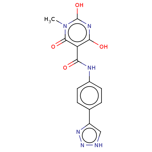 BDBM524201 US11168075, Compound Formula (Ig)
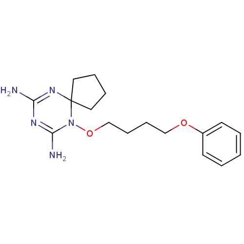 6,8,10-Traiazaspiro[4.5]deca-6,8-diene-7,9-diamine, Ig BDBM82111
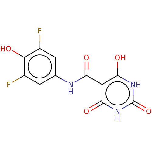
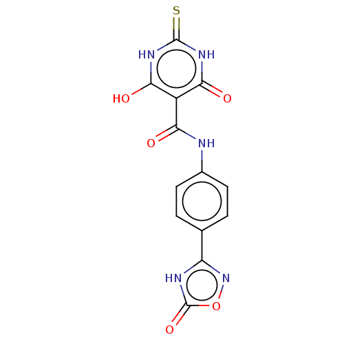 6-Hydroxy-4-oxo-N-(4-(5-oxo-4,5-dihydro-1,2,4-oxadiazol-3-yl)phenyl)-2-thioxo-1,2,3,4-tetrahydropyrimidine-5-carboxamide US10093658, Compound Formula (Ig) BDBM289118
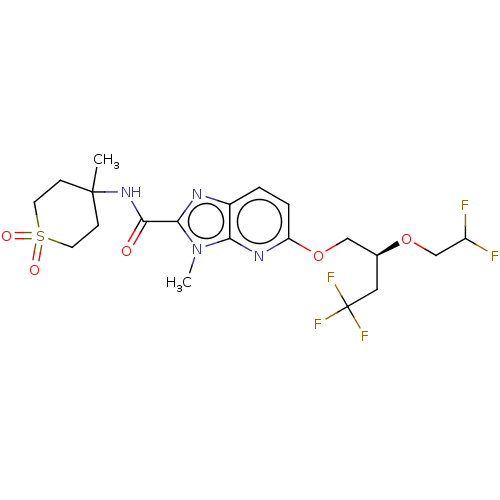 US20240254114, Example 63a BDBM689243 5-[2-(2,2-difluoroethoxy)-4,4,4-trifluoro- butoxy]-3-methyl-N-(4-methyl-1,1-dioxo- thian-4-yl)imidazo[4,5-b]pyridine-2- carboxamide (faster eluting on SFC IG column, 23%/77% MeOH/CO2)
- Zheng, S; Zhang, K; Zhang, X; Xiao, Y; Wang, T; Jiang, S Development of Inhibitors Targeting the V-Domain Ig Suppressor of T Cell Activation Signal Pathway. J Med Chem 65: 11900-11912 (2022)
- Wang, T; Wang, K; Zhang, Y; Zhang, K; Cai, S; Jiang, S; Xiao, Y; Zhang, X Novel Benzimidazoles as Potent Small-Molecule Inhibitors and Degraders of V-Domain Ig Suppressor of T-Cell Activation (VISTA). J Med Chem 66: 11881-11892 (2023)
- Chirkova, ZV; Kabanova, MV; Filimonov, SI; Abramov, IG; Petzer, A; Petzer, JP; Firgang, SI; Suponitsky, KY Bioorg Med Chem Lett 25: 1206-11 (2015)
- Boutselis, IG; Yu, X; Zhang, ZY; Borch, RF J Med Chem 50: 856-64 (2007)
- Cowell IG
- Birkinshaw, TN; Teague, SJ; Beech, C; Bonnert, RV; Hill, S; Patel, A; Reakes, S; Sanganee, H; Dougall, IG; Phillips, TT; Salter, S; Schmidt, J; Arrowsmith, EC; Carrillo, JJ; Bell, FM; Paine, SW; Weaver, R Bioorg Med Chem Lett 16: 4287-90 (2006)
- Ganley IG
- Smirnova, NA; Rakhman, I; Moroz, N; Basso, M; Payappilly, J; Kazakov, S; Hernandez-Guzman, F; Gaisina, IN; Kozikowski, AP; Ratan, RR; Gazaryan, IG Chem Biol 17: 380-91 (2010)
- Georg, IG; Ward, T; Faber, E; Hawkinson, JE; Wang, N; Schonbrunn, E US Patent US20240002339 (2024)
- Zheng, Z; Pinson, JA; Mountford, SJ; Orive, S; Schoenwaelder, SM; Shackleford, D; Powell, A; Nelson, EM; Hamilton, JR; Jackson, SP; Jennings, IG; Thompson, PE Eur J Med Chem 122: 339-351 (2016)
- Yoo, SK; Chung, JW; Jo, IG; Im, JH; Kang, KS; Kim, JY US Patent US9783551 (2017)
- Boby, ML; Fearon, D; Ferla, M; Filep, M; Koekemoer, L; Robinson, MC; null, null; Chodera, JD; Lee, AA; London, N; von Delft, A; von Delft, F; Achdout, H; Aimon, A; Alonzi, DS; Arbon, R; Aschenbrenner, JC; Balcomb, BH; Bar-David, E; Barr, H; Ben-Shmuel, A; Bennett, J; Bilenko, VA; Borden, B; Boulet, P; Bowman, GR; Brewitz, L; Brun, J; Bvnbs, S; Calmiano, M; Carbery, A; Carney, DW; Cattermole, E; Chang, E; Chernyshenko, E; Clyde, A; Coffland, JE; Cohen, G; Cole, JC; Contini, A; Cox, L; Croll, TI; Cvitkovic, M; De Jonghe, S; Dias, A; Donckers, K; Dotson, DL; Douangamath, A; Duberstein, S; Dudgeon, T; Dunnett, LE; Eastman, P; Erez, N; Eyermann, CJ; Fairhead, M; Fate, G; Fedorov, O; Fernandes, RS; Ferrins, L; Foster, R; Foster, H; Fraisse, L; Gabizon, R; García-Sastre, A; Gawriljuk, VO; Gehrtz, P; Gileadi, C; Giroud, C; Glass, WG; Glen, RC; Glinert, I; Godoy, AS; Gorichko, M; Gorrie-Stone, T; Griffen, EJ; Haneef, A; Hassell Hart, S; Heer, J; Henry, M; Hill, M; Horrell, S; Huang, QYJ; Huliak, VD; Hurley, MFD; Israely, T; Jajack, A; Jansen, J; Jnoff, E; Jochmans, D; John, T; Kaminow, B; Kang, L; Kantsadi, AL; Kenny, PW; Kiappes, JL; Kinakh, SO; Kovar, B; Krojer, T; La, VNT; Laghnimi-Hahn, S; Lefker, BA; Levy, H; Lithgo, RM; Logvinenko, IG; Lukacik, P; Macdonald, HB; MacLean, EM; Makower, LL; Malla, TR; Marples, PG; Matviiuk, T; McCorkindale, W; McGovern, BL; Melamed, S; Melnykov, KP; Michurin, O; Miesen, P; Mikolajek, H; Milne, BF; Minh, D; Morris, A; Morris, GM; Morwitzer, MJ; Moustakas, D; Mowbray, CE; Nakamura, AM; Neto, JB; Neyts, J; Nguyen, L; Noske, GD; Oleinikovas, V; Oliva, G; Overheul, GJ; Owen, CD; Pai, R; Pan, J; Paran, N; Payne, AM; Perry, B; Pingle, M; Pinjari, J; Politi, B; Powell, A; Pšenák, V; Pulido, I; Puni, R; Rangel, VL; Reddi, RN; Rees, P; Reid, SP; Reid, L; Resnick, E; Ripka, EG; Robinson, RP; Rodriguez-Guerra, J; Rosales, R; Rufa, DA; Saar, K; Saikatendu, KS; Salah, E; Schaller, D; Scheen, J; Schiffer, CA; Schofield, CJ; Shafeev, M; Shaikh, A; Shaqra, AM; Shi, J; Shurrush, K; Singh, S; Sittner, A; Sjö, P; Skyner, R; Smalley, A; Smeets, B; Smilova, MD; Solmesky, LJ; Spencer, J; Strain-Damerell, C; Swamy, V; Tamir, H; Taylor, JC; Tennant, RE; Thompson, W; Thompson, A; Tomásio, S; Tomlinson, CWE; Tsurupa, IS; Tumber, A; Vakonakis, I; van Rij, RP; Vangeel, L; Varghese, FS; Vaschetto, M; Vitner, EB; Voelz, V; Volkamer, A; Walsh, MA; Ward, W; Weatherall, C; Weiss, S; White, KM; Wild, CF; Witt, KD; Wittmann, M; Wright, N; Yahalom-Ronen, Y; Yilmaz, NK; Zaidmann, D; Zhang, I; Zidane, H; Zitzmann, N; Zvornicanin, SN Science 382: (2023)
- Vistnes, M; Christensen, G; Aronsen, M; Lunde, IG; Sjaastad, I; Carlson, CR US Patent US10322143 (2019)
- Madu, IG; Namanja, AT; Su, Y; Wong, S; Li, YJ; Chen, Y ACS Chem Biol 8: 1435-41 (2013)
- Carabet, LA; Lallous, N; Leblanc, E; Ban, F; Morin, H; Lawn, S; Ghaidi, F; Lee, J; Mills, IG; Gleave, ME; Rennie, PS; Cherkasov, A Eur J Med Chem 160: 108-119 (2018)
- Martínez-González, S; García, AB; Albarrán, MI; Cebriá, A; Amezquita-Alves, A; García-Campos, FJ; Martínez-Gago, J; Martínez-Torrecuadrada, J; Muñoz, IG; Blanco-Aparicio, C; Pastor, J Eur J Med Chem 201: (2020)
- Nair, MG; Nanavati, NT; Nair, IG; Kisliuk, RL; Gaumont, Y; Hsiao, MC; Kalman, TI J Med Chem 29: 1754-60 (1986)
- Chrunyk, BA; Rosner, MH; Cong, Y; McColl, AS; Otterness, IG; Daumy, GO Biochemistry 39: 7092-7099 (2000)
- Ahn, S; Ahn, M; Park, S; An, S; Park, IG; Hwang, SY; Gong, J; Oh, S; Jin, SH; Kim, HJ; Cheong, JH; Byun, Y; Noh, M Eur J Med Chem 245: (2023)
- Bano, S; Javed, K; Ahmad, S; Rathish, IG; Singh, S; Chaitanya, M; Arunasree, KM; Alam, MS Eur J Med Chem 65: 51-9 (2013)
- Safonov, IG; Heerding, DA; Keenan, RM; Price, AT; Erickson-Miller, CL; Hopson, CB; Levin, JL; Lord, KA; Tapley, PM Bioorg Med Chem Lett 16: 1212-6 (2006)
- Kaur, A; Piplani, S; Kaushik, D; Fung, J; Sakala, IG; Honda-Okubo, Y; Mehta, SK; Petrovsky, N; Salunke, DB RSC Med Chem 13: 622-637 (2022)
- Moreno-Cinos, C; Sassetti, E; Salado, IG; Witt, G; Benramdane, S; Reinhardt, L; Cruz, CD; Joossens, J; Van der Veken, P; Brötz-Oesterhelt, H; Tammela, P; Winterhalter, M; Gribbon, P; Windshügel, B; Augustyns, K J Med Chem 62: 774-797 (2019)
- Kick, EK; Busch, BB; Martin, R; Stevens, WC; Bollu, V; Xie, Y; Boren, BC; Nyman, MC; Nanao, MH; Nguyen, L; Plonowski, A; Schulman, IG; Yan, G; Zhang, H; Hou, X; Valente, MN; Narayanan, R; Behnia, K; Rodrigues, AD; Brock, B; Smalley, J; Cantor, GH; Lupisella, J; Sleph, P; Grimm, D; Ostrowski, J; Wexler, RR; Kirchgessner, T; Mohan, R ACS Med Chem Lett 7: 1207-1212 (2016)
- Boyd, HF; Fell, SC; Hickey, DM; Ife, RJ; Leach, CA; Macphee, CH; Milliner, KJ; Pinto, IL; Rawlings, DA; Smith, SA; Stansfield, IG; Stanway, SJ; Theobald, CJ; Whittaker, CM Bioorg Med Chem Lett 12: 51-5 (2001)
- Dale, R; Dong, S; Brocklehurst, D; Batchelor, AK; Simoes, MA; Thuillier, IG US Patent US12173301 (2024)
- Krasavin, M; Lukin, A; Bagnyukova, D; Zhurilo, N; Golovanov, A; Zozulya, S; Zahanich, I; Moore, D; Tikhonova, IG Eur J Med Chem 127: 357-368 (2017)
- ChEMBL_199992 (CHEMBL873249) Inhibitory activity against human chimeric L-selectin-Ig
- ChEMBL_200018 (CHEMBL810707) Inhibitory activity against human chimeric Selectin P-Ig
- ChEMBL_60884 (CHEMBL673531) Inhibitory activity against human chimeric E-selectin-Ig
- ChEMBL_217445 (CHEMBL823344) Inhibition of integrin alpha4-beta1 binding to VCAM-Ig
- ChEMBL_216159 (CHEMBL815330) Inhibition of MadCAM-Ig binding to alpha-4 beta-7 integrin
- ChEMBL_217618 (CHEMBL820361) Inhibitory activity of compound against alpha4-beta7 integrin using MAdCAM-Ig ligand
- ChEMBL_497066 (CHEMBL997702) Inhibition of recombinant LFA1/ICAM1-IG interaction by time-resolved fluorimetry method
- ChEMBL_535592 (CHEMBL987066) Inhibition of recombinant LFA1/ICAM1-IG interaction by time-resolved fluorimetry method
- ChEMBL_213405 (CHEMBL815428) Inhibition of I-VCAM-Ig binding to Very late antigen 4 (VLA-4)
- ChEMBL_214680 (CHEMBL818286) Inhibitory activity against very late antigen-4 (VLA-4) using VCAM-Ig ligand.
- ChEMBL_213378 (CHEMBL818161) Inhibition of [125I]VCAM-Ig binding to VLA-4 integrin of human Jurkat cells
- ChEMBL_213392 (CHEMBL818050) Inhibition of [125I]-VCAM-Ig binding to VLA-4 integrin of human Jurkat cells
- ChEMBL_213396 (CHEMBL818054) Inhibition of Very late antigen 4 (VLA-4) and VCAM-Ig fusion protein interaction
- ChEMBL_217476 (CHEMBL823552) Inhibition of [125I]VCAM-Ig binding to Alpha4-beta7 integrin of RPMI-8866 cells
- ChEMBL_217612 (CHEMBL819427) Inhibition of [125I]VCAM-Ig binding to Alpha4-beta7 integrin of RPMI-8866 cells
- ChEMBL_217613 (CHEMBL820356) Inhibition of [125I]VCAM-Ig binding to Alpha4-beta7 integrin of RPMI-8866 cells
- ChEMBL_1750186 (CHEMBL4184946) Inhibition of His-tagged PD-L1 binding to PD-1-Ig (unknown origin) preincubated with PD-L1 for 15 mins followed by PD-1-Ig addition measured after 15 mins by HTRF binding assay
- ChEMBL_213406 (CHEMBL815429) Inhibition of very late antigen-4 alpha4-beta1 (VLA-4), I-VCAM-Ig as radioligand
- ChEMBL_217605 (CHEMBL821055) Inhibition of [125I]MAdCAM-Ig binding to alpha4-beta7 integrin of human RPMI-8866 cells
- ChEMBL_217606 (CHEMBL818376) Inhibition of [125I]VCAM-Ig binding to alpha4-beta7 integrin of human RPMI-8866 cells
- ChEMBL_32534 (CHEMBL646568) Inhibition of [125I]MAdCAM-Ig binding to human integrin alpha4-beta7 of RPMI-8866 cells
- ChEMBL_213371 (CHEMBL817231) Inhibition of [125I]-VCAM-Ig binding to human integrin alpha4-beta7 receptor in RPMI-8866 cells
- ChEMBL_213373 (CHEMBL817233) In vitro inhibition of [125I]VCAM-Ig binding to very late antigen -4 on jurkat cells.
- ChEMBL_214674 (CHEMBL821412) Antagonistic activity against VLA-4 integrin of human jurkat cells using [125I]VCAM-Ig as radioligand
- ChEMBL_214678 (CHEMBL818284) Inhibition of [125I]VCAM-Ig binding to alpha4-beta1 (VLA-4) of human RPMI-8866 cells
- ChEMBL_214679 (CHEMBL818285) Inhibition of [125I]VCAM-Ig binding to Alpha-4 beta-1 (VLA-4) of Jurkat cells
- ChEMBL_215740 (CHEMBL823096) Inhibition of [125I]VCAM-Ig binding to human integrin alpha4-beta1 (VLA-4) of Jurkat cells
- ChEMBL_1667595 (CHEMBL4017391) Binding affinity to His-tagged PD-L1 (unknown origin) assessed as reduction in PD-L1/PD1-Ig interaction preincubated with PD-L1 for 15 mins followed by addition of PD1-Ig measured after 15 mins by HTRF assay
- ChEMBL_213389 (CHEMBL818047) Inhibition of [125I]VCAM-Ig binding to VLA-4 of Jurkat cells in the presence of [Ca2+]
- ChEMBL_213390 (CHEMBL818048) Inhibition of [125I]VCAM-Ig binding to VLA-4 of Jurkat cells in the presence of Mn2+
- ChEMBL_880326 (CHEMBL2216328) Inhibition of immobilized rhPSGL-Ig binding to soluble P-selectin by surface plasmon resonance competitive binding assay
- ChEMBL_1931330 (CHEMBL4434581) Inhibition of Ig-tagged human PD1/His-tagged human PDL1 interaction incubated for 30 mins by HTRF assay
- ChEMBL_2077183 (CHEMBL4732974) Displacement of 125I-VCAM-Ig from VLA4 in human Jurkat cells incubated for 30 mins by scintillation counting method
- ChEMBL_213372 (CHEMBL817232) Binding affinity towards VLA-4 (alpha4 beta-1) receptor in human Jurkat cells using [125I]VCAM-Ig as radioligand
- ChEMBL_610764 (CHEMBL1064399) Binding affinity to human N-terminal Ig-like domain D1D2 GPVI expressed in Escherichia coli DH5alpha by NMR spectroscopy
- ChEMBL_214677 (CHEMBL818283) Inhibitory activity against alpha-4 beta-1 receptor (VLA-4) in human RPMI-8866 cells using [125I]-VCAM-Ig ligand
- ChEMBL_215741 (CHEMBL823097) Binding affinity towards VLA-4 (alpha4 beta7) receptor in human RPMI-8866 B-cell line using [125I]MAdCAM-Ig as radioligand
- ChEMBL_215742 (CHEMBL823098) Binding affinity towards VLA-4 (alpha4 beta7) receptor in human RPMI-8866 B-cell line using [125I]MAdCAM-Ig as radioligand
- ChEMBL_217611 (CHEMBL819426) Inhibition of alpha4-beta7 integrin binding to radiolabeled vascular cell adhesion molecule-1 (VCAM-1) Ig fusion protein in human Jurkat cells
- ChEMBL_486011 (CHEMBL1017447) Inhibition of LFA1 expressed in human JY cells interaction with ICAM1-IG expressed in human HeLa cell monolayer after 45 mins by cell adhesion assay
- Homogenous Time-Resolved Fluorescence (HTRF) Assays Assays of Binding of Soluble PD-1 to Soluble PD-L1. Soluble PD-1 and soluble PD-L1 refers to proteins with carboxyl-end truncations that remove the transmembrane-spanning regions and are fused to heterologous sequences, specifically the Fc portion of the human immunoglobuling G sequence (Ig) or the hexahistidine epitope tag (His). All binding studies were performed in an HTRF assay buffer consisting of dPBS supplemented with 0.1% (w/v) bovine serum albumin and 0.05% (v/v) Tween-20. For the PD-1-Ig/PD-L1-His binding assay, inhibitors were pre-incubated with PD-L1-His (10 nM final) for 15m in 4 μl of assay buffer, followed by addition of PD-1-Ig (20 nM final) in 1 μl of assay buffer and further incubation for 15 m. PD-L1 fusion proteins from either human, cynomologous macaques, mouse, or other species were used. HTRF detection was achieved using europium crypate-labeled anti-Ig monoclonal antibody (1 nM final) and allophycocyanin (APC) labeled anti-His monoclonal antibody (20 nM final). Antibodies were diluted in HTRF detection buffer and 5 μl was dispensed on top of binding reaction. The reaction was allowed to equilibrate for 30 minutes and signal (665 nm/620 nm ratio) was obtained using an EnVision fluorometer. Additional binding assays were established between PD-1-Ig/PD-L2-His (20 and 5 nM, respectively), CD80-His/PD-L1-Ig (100 and 10 nM, respectively) and CD80-His/CTLA4-Ig (10 and 5 nM, respectively).
- ChEMBL_2241733 (CHEMBL5155943) Inhibition of PD-1-Ig/His-PD-L1 (unknown origin) protein-protein interaction incubated with PD-L1 for 15 mins followed by addition of PD-1 for 15 mins by HTRF assay
- ChEMBL_217307 (CHEMBL823928) Alpha4-beta1 integrin binding affinity was assessed by measuring the reduction in binding of [125I]VCAM-Ig to a suspension of Jurkat cells (a human T cell line alpha-4-beta-1-beta-7)
- ChEMBL_2327504 Inhibition of Ig-tagged PD-1 (unknown origin)/His-tagged PD-L1 (unknown origin) protein-protein interaction preincubated with PD-L1 for 15 mins followed by PD-1 addition measured after 15 mins by HTRF assay
- ChEMBL_217475 (CHEMBL823551) Alpha4-beta7 integrin binding affinity was determined in duplicate by a radioligand binding assay using [125I]-VCAM-Ig and a suspension of RPMI-8866 cells (a human B cell line alpha-4-beta-1-beta-7+)
- Homogenous Time-Resolved Fluorescence (HTRF) Binding Assay All binding studies were performed in an HTRF assay buffer consisting of dPBS supplemented with 0.1% (with) bovine serum albumin and 0.05% (v/v) Tween-20. For the h/PD-L1-His binding assay, inhibitors were pre-incubated with PD-L1-His (10 nM final) for 15 m in 4 μl of assay buffer, followed by addition of PD-1-Ig (20 nM final) in 1 μl of assay buffer and further incubation for 15 m. HTRF detection was achieved using europium crypate-labeled anti-Ig (1 nM final) and allophycocyanin (APC) labeled anti-His (20 nM final). Antibodies were diluted in HTRF detection buffer and 5 μl was dispensed on top of the binding reaction. The reaction mixture was allowed to equilibrate for 30 minutes and the resulting signal (665 nm/620 nm ratio) was obtained using an EnVision fluorometer. Additional binding assays were established between the human proteins PD-1-Ig/PD-L2-His (20 & 5 nM, respectively) and CD80-His/PD-L1-Ig (100 & 10 nM, respectively).
- ChEMBL_2332854 Inhibition of recombinant human C-terminal IG-tagged PD1 (25 to 167 residues) to human C-terminal His-tagged PD-L1 (18 to 239 residues) protein-protein interaction expressed in HEK-293T cells incubated for 15 mins by HTRF assay
- Homogenous Time-Resolved Fluorescence (HTRF) Binding Assay The interaction of PD-1 and PD-L1 can be assessed using soluble, purified preparations of the extracellular domains of the two proteins. The PD-1 and PD-L1 protein extracellular domains were expressed as fusion proteins with detection tags, for PD-1, the tag was the Fc portion of Immunoglobulin (PD-1-Ig) and for PD-L1 it was the 6 histidine motif (PD-L1-His). All binding studies were performed in an HTRF assay buffer consisting of dPBS supplemented with 0.1% (with) bovine serum albumin and 0.05% (v/v) Tween-20. For the h/PD-L1-His binding assay, inhibitors were pre-incubated with PD-L1-His (10 nM final) for 15m in 4 μl of assay buffer, followed by addition of PD-1-Ig (20 nM final) in 1 μ1 of assay buffer and further incubation for 15m. HTRF detection was achieved using europium crypate-labeled anti-Ig (1 nM final) and allophycocyanin (APC) labeled anti-His (20 nM final). Antibodies were diluted in HTRF detection buffer and 5 μl was dispensed on top of the binding reaction. The reaction mixture was allowed to equilibrate for 30 minutes and the resulting signal (665 nm/620 nm ratio) was obtained using an EnVision fluorometer. Additional binding assays were established between the human proteins PD-1-Ig/PD-L2-His (20 & 5 nM, respectively) and CD8O-His/PD-L1-Ig (100 & 10 nM, respectively). Recombinant Proteins: Human PD-1 (25-167) with a C-terminal human Fc domain of immunoglobulin G (Ig) epitope tag [hPD-1 (25-167)-3S-IG] and human PD-L1 (18-239) with a C-terminal His epitope tag [hPD-L1(18-239)-TVMV-His] were expressed in HEK293T cells and purified sequentially by ProteinA affinity chromatography and size exclusion chromatography. Human PD-L2-His and CD8O-His was obtained through commercial sources.
- Homogenous Time-Resolved Fluorescence (HTRF) Binding Assay The interaction of PD-1 and PD-L1 can be assessed using soluble, purified preparations of the extracellular domains of the two proteins. The PD-1 and PD-L1 protein extracellular domains were expressed as fusion proteins with detection tags, for PD-1, the tag was the Fc portion of Immunoglobulin (PD-1-Ig) and for PD-L1 it was the 6 histidine motif (PD-L1-His). All binding studies were performed in an HTRF assay buffer consisting of dPBS supplemented with 0.1% (with) bovine serum albumin and 0.05% (v/v) Tween-20. For the h/PD-L1-His binding assay, inhibitors were pre-incubated with PD-L1-His (10 nM final) for 15m in 4 μl of assay buffer, followed by addition of PD-1-Ig (20 nM final) in 1 μl of assay buffer and further incubation for 15 m. HTRF detection was achieved using europium crypate-labeled anti-Ig (1 nM final) and allophycocyanin (APC) labeled anti-His (20 nM final). Antibodies were diluted in HTRF detection buffer and 5 μl was dispensed on top of the binding reaction. The reaction mixture was allowed to equilibrate for 60 minutes and the resulting signal (665 nm/620 nm ratio) was obtained using an EnVision fluorometer. Additional binding assays were established between the human proteins PD-1-Ig/PD-L2-His (20 & 2 nM, respectively) and CD80-His/PD-L1-Ig (100 & 10 nM, respectively).Recombinant Proteins: Human PD-1 (25-167) with a C-terminal human Fc domain of immunoglobulin G (Ig) epitope tag [hPD-1 (25-167)-3S-IG] and human PD-L1 (18-239) with a C-terminal His epitope tag [hPD-L1(18-239)-TVMV-His] were expressed in HEK293T cells and purified sequentially by ProteinA affinity chromatography and size exclusion chromatography. Human PD-L2-His and CD80-His was obtained through commercial sources.
- Homogenous Time-Resolved Fluorescence (HTRF) Binding Assay The interaction of PD-1 and PD-L1 can be assessed using soluble, purified preparations of the extracellular domains of the two proteins. The PD-1 and PD-L1 protein extracellular domains were expressed as fusion proteins with detection tags, for PD-1, the tag was the Fc portion of Immunoglobulin (PD-1-Ig) and for PD-L1 it was the 6 histidine motif (PD-L1-His). All binding studies were performed in an HTRF assay buffer consisting of dPBS supplemented with 0.1% (with) bovine serum albumin and 0.05% (v/v) Tween-20. For the h/PD-L1-His binding assay, inhibitors were pre-incubated with PD-L1-His (10 nM final) for 15 m in 4 μl of assay buffer, followed by addition of PD-1-Ig (20 nM final) in 1 μl of assay buffer and further incubation for 15 m. HTRF detection was achieved using europium crypate-labeled anti-Ig (1 nM final) and allophycocyanin (APC) labeled anti-His (20 nM final). Antibodies were diluted in HTRF detection buffer and 5 μl was dispensed on top of the binding reaction. The reaction mixture was allowed to equilibrate for 30 minutes and the resulting signal (665 nm/620 nm ratio) was obtained using an EnVision fluorometer. Additional binding assays were established between the human proteins PD-1-Ig/PD-L2-His (20 & 5 nM, respectively) and CD80-His/PD-L1-Ig (100 & 10 nM, respectively).
- Homogenous Time-Resolved Fluorescence (HTRF) binding assay The interaction of PD-1 and PD-L1 can be assessed using soluble, purified preparations of the extracellular domains of the two proteins. The PD-1 and PD-L1 protein extracellular domains were expressed as fusion proteins with detection tags, for PD-1, the tag was the Fc portion of Immunoglobulin (PD-1-Ig) and for PD-L1 it was the 6 histidine motif (PD-L1-His). All binding studies were performed in an HTRF assay buffer consisting of dPBS supplemented with 0.1% (with) bovine serum albumin and 0.05% (v/v) Tween-20. For the h/PD-L1-His binding assay, inhibitors were pre-incubated with PD-L1-His (10 nM final) for 15 m in 4 μl of assay buffer, followed by addition of PD-1-Ig (20 nM final) in 1 μl of assay buffer and further incubation for 15 m. HTRF detection was achieved using europium crypate-labeled anti-Ig (1 nM final) and allophycocyanin (APC) labeled anti-His (20 nM final). Antibodies were diluted in HTRF detection buffer and 5 μl was dispensed on top of the binding reaction. The reaction mixture was allowed to equilibrate for 30 minutes and the resulting signal (665 nm/620 nm ratio) was obtained using an EnVision fluorometer. Additional binding assays were established between the human proteins PD-1-Ig/PD-L2-His (20 & 5 nM, respectively) and CD8O-His/PD-L1-Ig (100 & 10 nM, respectively).
- Homogenous Time-Resolved Fluorescence (HTRF) Binding Assay All binding studies were performed in an HTRF assay buffer consisting of dPBS supplemented with 0.1% (withv) bovine serum albumin and 0.05% (v/v) Tween-20. For the PD-1-Ig/PD-L1-His binding assay, inhibitors were pre-incubated with PD-L1-His (10 nM final) for 15 m in 4 μl of assay buffer, followed by addition of PD-1-Ig (20 nM final) in 1 μl of assay buffer and further incubation for 15 m. PD-L1 from either human, cyno, or mouse were used. HTRF detection was achieved using europium crypate-labeled anti-Ig (1 nM final) and allophycocyanin (APC) labeled anti-His (20 nM final). Antibodies were diluted in HTRF detection buffer and 5 μl was dispensed on top of binding reaction. The reaction mixture was allowed to equilibrate for 30 minutes and signal (665 nm/620 nm ratio) was obtained using an EnVision fluorometer. Additional binding assays were established between PD-1-Ig/PD-L2-His (20 & 5 nM, respectively), CD80-His/PD-L1-Ig (100 & 10 nM, respectively) and CD80-His/CTLA4-Ig (10 & 5 nM, respectively). Competition studies between biotinylated SEQ ID NO:71 and human PD-L1-His were performed as follows. Inhibitors were pre-incubated with PD-L1-His (10 nM final) for 60 m in 4 μl of assay buffer followed by addition of biotinylated SEQ ID NO:71 (0.5 nM final) in 1 μl of assay buffer. Binding was allowed to equilibrate for 30 m followed by addition of europium crypated labeled Strepatavidin (2.5 pM final) and APC-labeled anti-His (20 nM final) in 5 μl of HTRF buffer. The reaction was allowed to equilibrate for 30 m and signal (665 nm/620 nm ratio) was obtained using an EnVision fluorometer.
- Binding of PD-1 to PD-L1 Using Homogenous Time-Resolved Fluorescence (HTRF) Binding Assay Homogenous Time-Resolved Fluorescence (HTRF) Binding Assay. The interaction of PD-1 and PD-L1 can be assessed using soluble, purified preparations of the extracellular domains of the two proteins. The PD-1 and PD-L1 protein extracellular domains were expressed as fusion proteins with detection tags, for PD-1, the tag was the Fc portion of Immunoglobulin (PD-1-Ig) and for PD-L1 it was the 6 histidine motif (PD-L1-His). All binding studies were performed in an HTRF assay buffer consisting of dPBS supplemented with 0.1% (with) bovine serum albumin and 0.05% (v/v) Tween-20. For the h/PD-L1-His binding assay, inhibitors were pre-incubated with PD-L1-His (10 nM final) for 15 m in 4 μl of assay buffer, followed by addition of PD-1-Ig (20 nM final) in 1 μl of assay buffer and further incubation for 15 m. HTRF detection was achieved using europium cryptate-labeled anti-Ig (1 nM final) and allophycocyanin (APC) labeled anti-His (20 nM final). Antibodies were diluted in HTRF detection buffer and 5 μl was dispensed on top of the binding reaction. The reaction mixture was allowed to equilibrate for 30 minutes and the resulting signal (665 nm/620 nm ratio) was obtained using an EnVision fluorometer. Additional binding assays were established between the human proteins PD-1-Ig/PD-L2-His (20 & 5 nM, respectively) and CD80-His/PD-L1-Ig (100 & 10 nM, respectively).
- Homogenous Time-Resolved Fluorescence (HTRF) Assay TRF assay buffer consisting of dPBS supplemented with 0.1% (w/v) bovine serum albumin and 0.05% (v/v) Tween-20. For the PD-1-Ig/PD-L1-His binding assay, inhibitors were pre-incubated with PD-L1-His (10 nM final) for 15 m in 4 μl of assay buffer, followed by addition of PD-1-Ig (20 nM final) in 1 μl of assay buffer and further incubation for 15 m. PD-L1 fusion proteins from either human, cynomologous macaques, mouse, or other species were used. HTRF detection was achieved using europium crypate-labeled anti-Ig monoclonal antibody (1 nM final) and allophycocyanin (APC) labeled anti-His monoclonal antibody (20 nM final). Antibodies were diluted in HTRF detection buffer and 5 μl was dispensed on top of binding reaction. The reaction was allowed to equilibrate for 30 minutes and signal (665 nm/620 nm ratio) was obtained using an EnVision fluorometer.
- Homogenous Time-Resolved Fluorescence (HTRF) Binding Assay Homogenous Time-Resolved Fluorescence (HTRF) Assays of Binding of Soluble PD-1 to Soluble PD-L1. Soluble PD-1 and soluble PD-L1 refers to proteins with carboxyl-end truncations that remove the transmembrane-spanning regions and are fused to heterologous sequences, specifically the Fc portion of the human immunoglobulin G sequence (Ig) or the hexahistidine epitope tag (His). All binding studies were performed in an HTRF assay buffer consisting of dPBS supplemented with 0.1% (w/v) bovine serum albumin and 0.05% (v/v) Tween-20. For the PD-1-Ig/PD-L1-His binding assay, inhibitors were pre-incubated with PD-L1-His (10 nM final) for 15 m in 4 μl of assay buffer, followed by addition of PD-1-Ig (20 nM final) in 1 μl of assay buffer and further incubation for 15 m. PD-L1 fusion proteins from either human, cynomologous macaques, mouse, or other species were used. HTRF detection was achieved using europium crypate-labeled anti-Ig monoclonal antibody (1 nM final) and allophycocyanin (APC) labeled anti-His monoclonal antibody (20 nM final). Antibodies were diluted in HTRF detection buffer and 5 μl was dispensed on top of binding reaction. The reaction was allowed to equilibrate for 30 minutes and signal (665 nm/620 nm ratio) was obtained using an EnVision fluorometer. Additional binding assays were established between PD-1-Ig/PD-L2-His (20 and 5 nM, respectively), CD80-His/PD-L1-Ig (100 and 10 nM, respectively) and CD80-His/CTLA4-Ig (10 and 5 nM, respectively). Binding/competition studies between biotinylated Compound No. 71 and human PD-L1-His were performed as follows. Macrocyclic compound inhibitors were pre-incubated with PD-L1-His (10 nM final) for 60 minutes in 4 μl of assay buffer followed by addition of biotinylated Compound No. 71 (0.5 nM final) in 1 μl of assay buffer. Binding was allowed to equilibrate for 30 minutes followed by addition of europium crypated labeled Streptavidin (2.5 μM final) and APC-labeled anti-His (20 nM final) in 5 μl of HTRF buffer. The reaction was allowed to equilibrate for 30 m and signal (665 nm/620 nm ratio) was obtained using an EnVision fluorometer.
- Homogenous Time-Resolved Fluorescence (HTRF) Binding Assays The ability of the macrocyclic peptides of the present disclosure to bind to PD-L1 was investigated using a PD-1/PD-L1 Homogenous Time-Resolved Fluorescence (HTRF) binding assay.MethodsHomogenous Time-Resolved Fluorescence (HTRF) Assays of Binding of Soluble PD-1 to Soluble PD-L1. Soluble PD-1 and soluble PD-L1 refers to proteins with carboxyl-end truncations that remove the transmembrane-spanning regions and are fused to heterologous sequences, specifically the Fc portion of the human immunoglobuling G sequence (Ig) or the hexahistidine epitope tag (His). All binding studies were performed in an HTRF assay buffer consisting of dPBS supplemented with 0.1% (w/v) bovine serum albumin and 0.05% (v/v) Tween-20. For the PD-1-Ig/PD-L1-His binding assay, inhibitors were pre-incubated with PD-L1-His (10 nM final) for 15m in 4 μl of assay buffer, followed by addition of PD-1-Ig (20 nM final) in 1 μl of assay buffer and further incubation for 15 m. PD-L1 fusion proteins from either human, cynomologous macaques, mouse, or other species were used. HTRF detection was achieved using europium crypate-labeled anti-Ig monoclonal antibody (1 nM final) and allophycocyanin (APC) labeled anti-His monoclonal antibody (20 nM final). Antibodies were diluted in HTRF detection buffer and 5 μl was dispensed on top of binding reaction. The reaction was allowed to equilibrate for 30 minutes and signal (665 nm/620 nm ratio) was obtained using an EnVision fluorometer. Additional binding assays were established between PD-1-Ig/PD-L2-His (20 and 5 nM, respectively), CD8O-His/PD-L1-Ig (100 and 10 nM, respectively) and CD80-His/CTLA4-Ig (10 and 5 nM, respectively). Binding/competition studies between biotinylated Compound No. 71 and human PD-L1-His were performed as follows. Macrocyclic peptide inhibitors were pre-incubated with PD-L1-His (10 nM final) for 60 minutes in 4 μl of assay buffer followed by addition of biotinylated Compound No. 71 (0.5 nM final) in 1 μl of assay buffer. Binding was allowed to equilibrate for 30 minutes followed by addition of europium crypated labeled Streptavidin (2.5 pM final) and APC-labeled anti-His (20 nM final) in 5 μl of HTRF buffer. The reaction was allowed to equilibrate for 30 m and signal (665 nm/620 nm ratio) was obtained using an EnVision fluorometer.
- PD-1/PD-L1 Homogenous Time-Resolved Fluorescence (HTRF) binding assay Homogenous Time-Resolved Fluorescence (HTRF) Assays of Binding of Soluble PD-1 to Soluble PD-L1. Soluble PD-1 and soluble PD-L1 refers to proteins with carboxyl-end truncations that remove the transmembrane-spanning regions and are fused to heterologous sequences, specifically the Fc portion of the human immunoglobuling G sequence (Ig) or the hexahistidine epitope tag (His). All binding studies were performed in an HTRF assay buffer consisting of dPBS supplemented with 0.1% (w/v) bovine serum albumin and 0.05% (v/v) Tween-20. For the PD-1-Ig/PD-L1-His binding assay, inhibitors were pre-incubated with PD-L1-His (10 nM final) for 15 m in 4 μl of assay buffer, followed by addition of PD-1-Ig (20 nM final) in 1 μl of assay buffer and further incubation for 15 m. PD-L1 fusion proteins from either human, cynomologous macaques, mouse, or other species were used. HTRF detection was achieved using europium crypate-labeled anti-Ig monoclonal antibody (1 nM final) and allophycocyanin (APC) labeled anti-His monoclonal antibody (20 nM final). Antibodies were diluted in HTRF detection buffer and 5 μl was dispensed on top of binding reaction. The reaction was allowed to equilibrate for 30 minutes and signal (665 nm/620 nm ratio) was obtained using an EnVision fluorometer. Additional binding assays were established between PD-1-Ig/PD-L2-His (20 and 5 nM, respectively), CD80-His/PD-L1-Ig (100 and 10 nM, respectively) and CD80-His/CTLA4-Ig (10 and 5 nM, respectively). Binding/competition studies between biotinylated Compound No. 71 and human PD-L1-His were performed as follows. Macrocyclic peptide inhibitors were pre-incubated with PD-L1-His (10 nM final) for 60 minutes in 4 μl of assay buffer followed by addition of biotinylated Compound No. 71 (0.5 nM final) in 1 μl of assay buffer. Binding was allowed to equilibrate for 30 minutes followed by addition of europium crypated labeled Streptavidin (2.5 pM final) and APC-labeled anti-His (20 nM final) in 5 μl of HTRF buffer. The reaction was allowed to equilibrate for 30 m and signal (665 nm/620 nm ratio) was obtained using an EnVision fluorometer.
- ChEMBL_2327512 Inhibition of C-terminal 3S-IG-tagged recombinant human PD-1 (25 to 167 residues)/C-terminal 6xHis-tagged recombinant human PD-L1 (18 to 239 residues) expressed in HEK293T cells protein-protein interaction preincubated with PD-L1 for 15 mins followed by PD-1 addition measured after 15 mins by HTRF assay
- ChEMBL_1988151 (CHEMBL4621698) Inhibition of human C-terminal Ig-Fc-tagged PD1 (Leu25 to Gln167 residues) expressed in HEK293 cells/human C-terminal epitope-His-tagged PDL1 (Phe19 to Arg238 residues) expressed in HEK293 cells protein-protein interaction preincubated for 15 mins with PDL1 followed by PD1 addition and measured after 15 mins by APC-labeled anti-His antibody/Eu-labeled anti-human IgG based HTRF assay
- In Vitro Agonist Binding Assay Preparation of Membrane Fractions from CHO-h5-HT1A Cells. Membranes from CHO cells stably expressing the human 5-HT1A receptor at a density of 8 pmol/mg membrane protein with 5-HT2c receptor background were prepared. Cells were grown in DMEM/F-12 medium supplemented with 5% fatal bovine serum, 50 ig/mL geneticin, and 50 ig/mL hygromycin B in a humidified atmosphere of 5% CO2 until they reached confluence. Cells were harvested by centrifugation (800 g for 5 min) and homogenized using a polytron homogenizer (Polytron, CH-6010 Kreiens-Lu, Brinkman Instrument, Westbury, N.Y.) in buffer containing 20 mM HEPES, pH7.4, 3 mM MgCl2, and a cocktail of protease inhibitors (Sigma-Aldrich, St. Louis, Mo.) at 1:2000 dilution. The homogenate was centrifuged (Beckman Optima LE80K Ultracentrifuge) at 100 000 g for 15 min at 4° C. The pellet was suspended in the same buffer and recentrifuged. The final pellet was suspended in assay buffer containing 20 mM HEPES, pH 7.4, 3 mM MgCl2, 100 mM NaCl, and a mixture of protease inhibitors and stored at -70° C. Protein concentration was determined by detergent compatible colorimetric assay using DC Protein Assay Reagents as recommended by the manufacturer (Bio-Rad, Hercules, Calif.).
- Binding Assay The inhibition assay to screen for and characterize glycomimetic antagonists of E-selectin is a competitive binding assay, which allows the determination of IC50 values. E-selectin/Ig chimera was immobilized in 96 well microtiter plates by incubation at 37° C. for 2 hours. To reduce nonspecific binding, bovine serum albumin was added to each well and incubated at room temperature for 2 hours. The plate was washed and serial dilutions of the test compounds were added to the wells in the presence of conjugates of biotinylated, sLea polyacrylamide with streptavidin/horseradish peroxidase and incubated for 2 hours at room temperature.To determine the amount of sLea bound to immobilized E-selectin after washing, the peroxidase substrate, 3,3′,5,5′ tetramethylbenzidine (TMB) was added. After 3 minutes, the enzyme reaction was stopped by the addition of H3PO4, and the absorbance of light at a wavelength of 450 nm was determined.
- Biological Assay Assay buffer was 50 mM Tris pH 7.5 containing 1 mM EGTA (ethylene glycol tetraacetic acid), 1 mM DTT (dithiothreitol), 0.1 mM Na3VO4, 5 mM MgCl2, 0.01% Tween 20. Assays were carried out in 384 well Mesoscale high binding plates which had been coated with myelin basic protein (MBP) and blocked with bovine serum albumin to prevent non-specific protein binding. All compounds tested were dissolved in dimethyl sulfoxide (DMSO) and further dilutions were made in assay buffer. Final DMSO concentration was 1% (v/v) in assays. Incubations consisted of compound (1% DMSO in control and blank wells), 25 μM Adenosine-5′-triphosphate (ATP), and 10 nM NIK/MAP3K14 substituting enzyme with buffer in the blank wells. Incubations were carried out for 1 h at 25° C. and were followed by washing and sequential incubation with rabbit anti-phospho-MBP and anti-rabbit Ig Sulfotag antibody before reading bound Sulfotag on a Mesoscale Discovery. Signal obtained in the wells containing blank samples was subtracted from all other wells and IC50's were determined by fitting a sigmoidal curve to % inhibition of control versus Log10 compound concentration.
- E-Selectin Activity - Binding Assay The inhibition assay to screen for and characterize glycomimetic antagonists of E-selectin is a competitive binding assay, which allows the determination of IC50 values. E-selectin/Ig chimera was immobilized in 96 well microtiter plates by incubation at 37° C. for 2 hours. To reduce nonspecific binding, bovine serum albumin was added to each well and incubated at room temperature for 2 hours. The plate was washed, and serial dilutions of the test compounds were added to the wells in the presence of conjugates of biotinylated, sLea polyacrylamide with streptavidin/horseradish peroxidase and incubated for 2 hours at room temperature.To determine the amount of sLea bound to immobilized E-selectin after washing, the peroxidase substrate, 3,3′,5,5′ tetramethylbenzidine (TMB) was added. After 3 minutes, the enzyme reaction was stopped by the addition of H3PO4, and the absorbance of light at a wavelength of 450 nm was determined. The concentration of test compound required to inhibit binding by 50% was determined and reported as the IC50 value for each glycomimetic E-selectin antagonist as shown in the table below.
- TR-FRET Assay Protein arginine methyltransferase 5 (PRMT5) is a type II arginine methyltransferase that catalyze mono- and symmetric demethylation on arginine residues of histone or non-histone proteins in presence of S-adenosylmethionine (AdoMet or SAM) a cofactor responsible for donating the methyl group. PRMT5 is reported to be overexpressed in several human cancers. To identify compounds that inhibit the PRMT5 and decrease its activity, a TR-FRET based assay has been established. Time-resolved fluorescence resonance energy transfer (TR-FRET) HTS assays are homogeneous proximity assays where the interaction of two dye-labeled binding partners is detected by the energy transfer between a donor and an acceptor dye, and the subsequent light emission by the acceptor dye. PRMT5 catalyzes Histone H4 peptide [1-16] which is biotin tagged to the Lysine amino acid at carboxyl end, in presence of S-adenosyl-1-methionine (SAM) to methylate the peptide. The antibody specific to mono methylated H4 peptide (H4R3) with Ig conjugate binds to the methylated peptide, indirectly binding to the Europium lanthanide. SureLight Allophycocyanin-Streptavidin binds to the biotin tag of the peptide, therefore accepting the energy transferred from the Europium lanthanide. This energy transfer between Europium to SureLight Allophycocyanin is a direct measure of the activity/inhibition of the PRMT5 enzyme.
- PRMT5 Inhibition Based on TR-FRET Assay Protein arginine methyltransferase 5 (PRMT5) is a type II arginine methyltransferase that catalyze mono- and symmetric demethylation on arginine residues of histone or non-histone proteins in presence of S-adenosylmethionine (AdoMet or SAM) a cofactor responsible for donating the methyl group. PRMT5 is reported to be overexpressed in several human cancers. To identify compounds that inhibit the PRMT5 and decrease its activity, a TR-FRET based assay has been established. Time-resolved fluorescence resonance energy transfer (TR-FRET) HTS assays are homogeneous proximity assays where the interaction of two dye-labeled binding partners is detected by the energy transfer between a donor and an acceptor dye and the subsequent light emission by the acceptor dye. PRMT5 catalyzes Histone H4 peptide [1-16] which is biotin tagged to the Lysine amino acid at carboxyl end, in presence of S-adenosyl-1-methionine (SAM) to methylate the peptide. The antibody specific to mono methylated H4 peptide (H4R3) with Ig conjugate binds to the methylated peptide, indirectly binding to the Europium lanthanide. SureLight Allophycocyanin-Streptavidin binds to the biotin tag of the peptide, therefore accepting the energy transferred from the Europium lanthanide. This energy transfer between Europium to SureLight Allophycocyanin is a direct measure of the activity/inhibition of the PRMT5 enzyme.
- Inhibit Human Recombinant Assay SSAO/VAP-1: Human recombinant SSAO/VAP-1 amine oxidase activity was determined using the coupled colorimetric method as described for monoamine oxidase, copper-containing amine oxidases and related enzymes (Holt A. and Palcic M., A peroxidase-coupled continuous absorbance plate-reader assay for flavin monoamine oxidases, copper-containing amine oxidases and related enzymes. Nat Protoc 2006; 1: 2498-2505). Briefly, a cloned cDNA template corresponding to residues 34-763 of human SSAO/VAP-1, and incorporating a mouse Ig kappa (κ) signal sequence, N-terminal flag epitope tag and tobacco etch virus (TEV) cleavage site, was assembled in a mammalian expression vector (pLO-CMV) by Geneart AG. This vector containing human SSAO/VAP-1 residues was transfected into CHO-K1 glycosylation mutant cell line, Lec 8. A clone stably expressing human SSAO/VAP-1 was isolated and cultured in large scale. Active human SSAO/VAP-1 was purified and recovered using immunoaffinity chromatography. This was used as the source for SSAO/VAP-1 activity. A high-throughput colorimetric assay was developed using either 96 or 384 well format. Briefly, in a standard 96 well plate assay 50 μL of purified human SSAO/VAP-1 (0.25 μg/mL) in 0.1 M sodium phosphate buffer (pH 7.4) was added into each well. Test compounds were dissolved in DMSO and tested in a Concentration Response Curve (CRC) with 4-11 data points, typically in the micromolar or nanomolar range after incubation with human SSAO/VAP-1 for 30 min at 37° C.
- SSAO/VAP-1 Amine Oxidase Activity Assay Briefly, a cloned cDNA template corresponding to residues 34-763 of human SSAO/VAP-1, and incorporating a mouse Ig kappa (κ) signal sequence, N-terminal flag epitope tag and tobacco etch virus (TEV) cleavage site, was assembled in a mammalian expression vector (pLO-CMV) by Geneart AG. This vector containing human SSAO/VAP-1 residues was transfected into CHO-Kl glycosylation mutant cell line, Lec 8. A clone stably expressing human SSAO/VAP-1 was isolated and cultured in large scale. Active human SSAO/VAP-1 was purified and recovered using immunoaffinity chromatography. This was used as the source for SSAO/VAP-1 activity. A high-throughput colorimetric assay was developed using either 96 or 384 well format. Briefly, in a standard 96 well plate assay 50 μL of purified human SSAO/VAP-1 (0.25 μg/mL) in 0.1 M sodium phosphate buffer (pH 7.4) was added into each well. Test compounds were dissolved in DMSO and tested in a Concentration Response Curve (CRC) with 4-11 data points, typically in the micromolar or nanomolar range after incubation with human SSAO/VAP-1 for 30 min at 37° C. After 30 min incubation, 50 μL of the reaction mixture containing 600 μM benzylamine (Sigma Aldrich), 120 μM Amplex Red (Sigma Aldrich) and 1.5 U/mL horseradish peroxidase (Sigma Aldrich) prepared in 0.1 M sodium phosphate buffer (pH 7.4) were added to the corresponding well. The fluorescence unit (RFU) was read every 2.5 min for 30 min at 37° C. excitation 565 nm and emission 590 (Optima; BMG labtech).
- PARG Activity Assay PARG Activity Assay was performed in Streptavidin-coated 96-well plates. Terminal-biotinylated poly (ADP-ribose) polymer was diluted to a final concentration of 25 nM with 0.2% bovine serum albumin (BSA) containing phosphate-buffered saline solution (PBS) and used for the substrate solution. The substrate solution was added to the 96-well plate at the volume of 200 micro liter per well and incubated for 3 hours at room temperature. After the incubation, the substrate solution was discarded and assay plate was washed 4 times with 220 micro liter of PBS containing 0.1% Tween20 (PBST). Recombinant human PARG protein was diluted with 0.2% BSA containing PBS to a final concentration of 0.0185 nM. The diluted enzyme solution was pre-incubated with compounds for 1 hour at room temperature in a 96-well plate and transferred 100 micro liter per well to the poly (ADP-ribose) substrate-coated assay plate. The plate was incubated for 1 hour at 25° C. and then washed 4 times with 220 micro liters of PBST. For the detection of non-digested poly (ADP-ribose) polymer on the plate, mouse anti-poly (ADP-ribose) antibody diluted with 0.2% BSA containing PBS was added 100 micro liters per well and incubated 1 hour at room temperature. Plate was washed 4 times with TBST and 100 micro liter of HRP-labeled anti-mouse Ig antibody was added to the wells. Plate was incubated for 30 minutes at room temperature and then washed 4 times with PBST. For the detection 100 micro liters of TMB substrate solution was added to the wells. After sufficient color development, reaction was stopped by addition of 50 micro liter of 0.2 M Sulfonic acid.
- Inhibit Human Recombinant Assay Human recombinant SSAO/VAP-1 amine oxidase activity was determined using the coupled colorimetric method as described for monoamine oxidase, copper-containing amine oxidases and related enzymes (Holt A. and Palcic M., A peroxidase-coupled continuous absorbance plate-reader assay for flavin monoamine oxidases, copper-containing amine oxidases and related enzymes. Nat Protoc 2006; 1: 2498-2505). Briefly, a cloned cDNA template corresponding to residues 34-763 of human SSAO/VAP-1, and incorporating a mouse Ig kappa (κ) signal sequence, N-terminal flag epitope tag and tobacco etch virus (TEV) cleavage site, was assembled in a mammalian expression vector (pLO-CMV) by Geneart AG. This vector containing human SSAO/VAP-1 residues was transfected into CHO-K1 glycosylation mutant cell line, Lec 8. A clone stably expressing human SSAO/VAP-1 was isolated and cultured in large scale. Active human SSAO/VAP-1 was purified and recovered using immunoaffinity chromatography. This was used as the source for SSAO/VAP-1 activity. A high-throughput colorimetric assay was developed using either 96 or 384 well format. Briefly, in a standard 96 well plate assay 50 μL of purified human SSAO/VAP-1 (0.25 μg/mL) in 0.1 M sodium phosphate buffer (pH 7.4) was added into each well. Test compounds were dissolved in DMSO and tested in a Concentration Response Curve (CRC) with 4-11 data points, typically in the micromolar or nanomolar range after incubation with human SSAO/VAP-1 for 30 min at 37° C. After 30 min incubation, 50 μL of the reaction mixture containing 600 μM benzylamine (Sigma Aldrich), 120 μM Amplex Red (Sigma Aldrich) and 1.5 U/mL horseradish peroxidase (Sigma Aldrich) prepared in 0.1 M sodium phosphate buffer (pH 7.4) were added to the corresponding well. The fluorescence unit (RFU) was read every 2.5 min for 30 min at 37° C. excitation 565 nm and emission 590 (Optima; BMG labtech).
- Inhibition Assay Human recombinant SSAO/VAP-1 amine oxidase activity was determined using the coupled colorimetric method as described for monoamine oxidase, copper-containing amine oxidases and related enzymes (Holt A. and Palcic M., A peroxidase-coupled continuous absorbance plate-reader assay for flavin monoamine oxidases, copper-containing amine oxidases and related enzymes. Nat Protoc 2006; 1: 2498-2505). Briefly, a cloned cDNA template corresponding to residues 34-763 of human SSAO/VAP-1, and incorporating a mouse Ig kappa (κ) signal sequence, N-terminal flag epitope tag and tobacco etch virus (TEV) cleavage site, was assembled in a mammalian expression vector (pLO-CMV) by Geneart AG. This vector containing human SSAO/VAP-1 residues was transfected into CHO-K1 glycosylation mutant cell line, Lee 8. A clone stably expressing human SSAO/VAP-1 was isolated and cultured in large scale. Active human SSAO/VAP-1 was purified and recovered using immunoaffinity chromatography. This was used as the source for SSAO/VAP-1 activity. A high-throughput colorimetric assay was developed using either 96 or 384 well format. Briefly, in a standard 96 well plate assay 50 μL of purified human SSAO/VAP-1 (0.25 μg/mL) in 0.1 M sodium phosphate buffer (pH 7.4) was added into each well. Test compounds were dissolved in DMSO and tested in a Concentration Response Curve (CRC) with 4-11 data points, typically in the micromolar or nanomolar range after incubation with human SSAO/VAP-1 for 30 min at 37° C. After 30 min incubation, 50 L of the reaction mixture containing 600 μM benzylamine (Sigma Aldrich), 120 M Amplex Red (Sigma Aldrich) and 1.5 U/mL horseradish peroxidase (Sigma Aldrich) prepared in 0.1 M sodium phosphate buffer (pH 7.4) were added to the corresponding well. The fluorescence unit (RFU) was read every 2.5 min for 30 min at 37° C. excitation 565 nm and emission 590