 BDBM82095 PGE2 13,14-dihydro
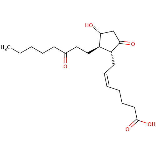 BDBM82093 PGE2 13,14-dihydro CAS_363-23-5 13,14-DIHYDRO-15-KETO PROSTAGLANDIN E2 13,14-dihydro-15-oxo-prostaglandin E2 PGE2, 13,14-dihydro15-oxo
 (Z)-7-[(1R,2R,3R)-3-Hydroxy-2-((E)-(R)-3-hydroxy-oct-1-enyl)-5-oxo-cyclopentyl]-hept-5-enoic acid CHEMBL64804 BDBM50101822 US9180116, PGE2 PGE2, 15-epi
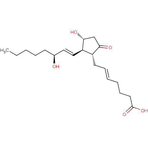 BDBM50213982 PGE2, 5,6-trans (E)-7-((1R,2R,3R)-3-hydroxy-2-((S,E)-3-hydroxyoct-1-enyl)-5-oxocyclopentyl)hept-5-enoic acid
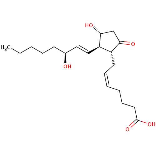 BDBM35847 [3H]Prostaglandin E2 PGE2 CHEMBL548 DINOPROSTONE (15S)-prostaglandin E2 prostaglandin E2 [3H]PGE2 (E,Z)-(1R,2R,3R)-7-(3-Hydroxy-2-((3S)-(3-hydroxy-1-octenyl))-5-oxocyclopentyl)-5-heptenoic acid [3H]Dinoprostone (5Z,11alpha,13E,15S)-11,15-dihydroxy-9-oxoprosta-5,13-dien-1-oic acid (5Z,13E,15S)-11alpha,15-dihydroxy-9-oxoprosta-5,13-dien-1-oic acid
- Boss, C; Corminboeuf, O; Fretz, H; Lyothier, I; Pozzi, D; Richard-Bildstein, S; Siendt, H; Sifferlen, T Pyrimidine derivatives as PGE2 receptor modulators US Patent US11839613 (2023)
- Fretz, H; Lyothier, I; Pothier, J; Richard-Bildstein, S; Sifferlen, T; Wyder Peters, L; Pozzi, D; Corminboeuf, O N-substituted indole derivatives as PGE2 receptor modulators US Patent US12011444 (2024)
- Bachovchin, WW; Lai, H; Wu, W Combination therapies using immuno-dash inhibitors and PGE2 antagonists US Patent US11096924 (2021)
- Bachovchin, WW; Lai, H; Wu, W Combination therapies using caspase-1 dependent anticancer agents and PGE2 antagonists US Patent US11559537 (2023)
- Smith, B; Chang, HH; Medda, F; Gokhale, V; Dietrich, J; Davis, A; Meuillet, EJ; Hulme, C Synthesis and biological activity of 2-aminothiazoles as novel inhibitors of PGE2 production in cells. Bioorg Med Chem Lett 22: 3567-70 (2012)
- Zhao, Z; Araldi, GL; Xiao, Y; Reddy, AP; Liao, Y; Karra, S; Brugger, N; Fischer, D; Palmer, E Synthesis and evaluation of novel pyrazolidinone analogs of PGE2 as EP2 and EP4 receptors agonists. Bioorg Med Chem Lett 17: 6572-5 (2007)
- Kambe, T; Maruyama, T; Nakai, Y; Oida, H; Maruyama, T; Abe, N; Nishiura, A; Nakai, H; Toda, M Synthesis and evaluation of¿-lactam analogs of PGE2 as EP4 and EP2/EP4 agonists. Bioorg Med Chem 20: 3502-22 (2012)
- Xiao, Y; Araldi, GL; Zhao, Z; Brugger, N; Karra, S; Fischer, D; Palmer, E Discovery of novel prostaglandin analogs of PGE2 as potent and selective EP2 and EP4 receptor agonists. Bioorg Med Chem Lett 17: 4323-7 (2007)
- Medda, F; Sells, E; Chang, HH; Dietrich, J; Chappeta, S; Smith, B; Gokhale, V; Meuillet, EJ; Hulme, C Synthesis and biological activity of aminophthalazines and aminopyridazines as novel inhibitors of PGE2 production in cells. Bioorg Med Chem Lett 23: 528-31 (2012)
- Cameron, KO; Lefker, BA; Ke, HZ; Li, M; Zawistoski, MP; Tjoa, CM; Wright, AS; DeNinno, SL; Paralkar, VM; Owen, TA; Yu, L; Thompson, DD Discovery of CP-533536: an EP2 receptor selective prostaglandin E2 (PGE2) agonist that induces local bone formation. Bioorg Med Chem Lett 19: 2075-8 (2009)
- Kassab, SE; Khedr, MA; Ali, HI; Abdalla, MM Discovery of new indomethacin-based analogs with potentially selective cyclooxygenase-2 inhibition and observed diminishing to PGE2 activities. Eur J Med Chem 141: 306-321 (2017)
- Kang, SM; Lee, J; Jin, JH; Kim, M; Lee, S; Lee, HH; Shin, JS; Lee, KT; Lee, JY Synthesis and PGE2 production inhibition of s-triazine derivatives as a novel scaffold in RAW 264.7 macrophage cells. Bioorg Med Chem Lett 24: 5418-22 (2015)
- Yang, Z; Truong, TN; Pham, TA; Lee, JW; Kim, SS; Park, H Synthesis of 1,5-diarylhaloimidazole analogs and their inhibitory activities against PGE2 production from LPS-treated RAW 264.7 cells. Bioorg Med Chem 20: 6256-9 (2012)
- Kim, M; Lee, S; Park, EB; Kim, KJ; Lee, HH; Shin, JS; Fischer, K; Koeberle, A; Werz, O; Lee, KT; Lee, JY Hit-to-lead optimization of phenylsulfonyl hydrazides for a potent suppressor of PGE2 production: Synthesis, biological activity, and molecular docking study. Bioorg Med Chem Lett 26: 94-9 (2015)
- ChEMBL_467683 (CHEMBL937605) Displacement of [3H]PGE2 from human PGE2-EP1 receptor expressed in CHO-K1 cells
- ChEMBL_467689 (CHEMBL937611) Antagonist activity at PGE2-EP1 receptor assessed as PGE2-induced response by schild analysis
- ChEMBL_437304 (CHEMBL906705) Inhibition of ovine COX1 by measuring PGE2
- ChEMBL_468124 (CHEMBL934144) Displacement of [3H]PGE2 from EP1 receptor
- ChEMBL_544157 (CHEMBL1013438) Displacement of [3H]PGE2 from EP1 receptor
- ChEMBL_437305 (CHEMBL906707) Inhibition of human recombinant COX2 by measuring PGE2
- ChEMBL_468777 (CHEMBL931917) Displacement of [3H]PGE2 from human EP2 receptor
- ChEMBL_468798 (CHEMBL932038) Displacement of [3H]PGE2 from human EP1 receptor
- ChEMBL_468799 (CHEMBL932039) Displacement of [3H]PGE2 from human EP3 receptor
- ChEMBL_515713 (CHEMBL993101) Displacement of [3H]PGE2 from human EP3 receptor
- ChEMBL_558515 (CHEMBL963081) Displacement of [3H]PGE2 from human EP3 receptor
- ChEMBL_558524 (CHEMBL963090) Displacement of [3H]PGE2 from mouse EP3 receptor
- ChEMBL_618650 (CHEMBL1101699) Displacement of [3H]PGE2 from human EP3 receptor
- ChEMBL_2263055 (CHEMBL5218066) Inhibition of 15-PGDH (unknown origin) using PGE2 as substrate
- ChEMBL_547430 (CHEMBL1026720) Inhibition of bovine seminal microsomal COX1 assessed as PGE2 production
- ChEMBL_547431 (CHEMBL1026721) Inhibition of sheep placental cotyledons COX2 assessed as PGE2 production
- ChEMBL_626787 (CHEMBL1107234) Displacement of [3H]PGE2 from human EP3 receptor in buffer
- ChEMBL_836140 (CHEMBL2077576) TP_TRANSPORTER: inhibition of PGE2 uptake in PGT-expressing HeLa cells
- ChEMBL_836787 (CHEMBL2075312) TP_TRANSPORTER: inhibition of PGE2 uptake in PGT-expressing HeLa cells
- ChEMBL_836797 (CHEMBL2075322) TP_TRANSPORTER: inhibition of PGE2 uptake in PGT-expressing HeLa cells
- ChEMBL_837082 (CHEMBL2076334) TP_TRANSPORTER: inhibition of PGE2 uptake in PGT-expressing HeLa cells
- ChEMBL_838709 (CHEMBL2078491) TP_TRANSPORTER: inhibition of PGE2 uptake in PGT-expressing HeLa cells
- ChEMBL_2193804 (CHEMBL5106164) Inhibition of human mPGES-1 assessed as reduction in PGE2 production
- ChEMBL_2375719 Inhibition of COX-2 (unknown origin) assessed as reduction in PGE2 production
- ChEMBL_2375720 Inhibition of COX-1 (unknown origin) assessed as reduction in PGE2 production
- ChEMBL_2447646 Inhibition of COX-2 (unknown origin) assessed as reduction in PGE2 production
- ChEMBL_473084 (CHEMBL921796) Inhibition of human COX2 mediated PGE2 production in human whole blood
- ChEMBL_789632 (CHEMBL1924717) Inhibition of COX2-mediated PGE2 production in human whole blood assay
- ChEMBL_456218 (CHEMBL888228) Displacement of [3H]PGE2 from human EP1 receptor expressed in HEK293 cells
- ChEMBL_456219 (CHEMBL888229) Displacement of [3H]PGE2 from human EP2 receptor expressed in HEK293 cells
- ChEMBL_456221 (CHEMBL888231) Displacement of [3H]PGE2 from human EP3 receptor expressed in HEK293 cells
- ChEMBL_456222 (CHEMBL888232) Displacement of [3H]PGE2 from human EP4 receptor expressed in HEK293 cells
- ChEMBL_477817 (CHEMBL931249) Displacement of [3H]PGE2 from human EP1 receptor expressed in CHOK1 cells
- ChEMBL_500469 (CHEMBL1009646) Displacement of [3H]PGE2 from human EP1 receptor expressed in CHO cells
- ChEMBL_625948 (CHEMBL1103467) Inhibition of sheep placental cotyledens COX2 assessed as PGE2 level by EIA
- ChEMBL_625949 (CHEMBL1103468) Inhibition of ram seminal vesicle COX1 assessed as PGE2 level by EIA
- ChEMBL_775624 (CHEMBL1913308) Displacement of [3H]-PGE2 from human EP4 receptor expressed in HEK293 cells
- ChEMBL_775625 (CHEMBL1913309) Displacement of [3H]-PGE2 from human EP2 receptor expressed in HEK293 cells
- ChEMBL_821751 (CHEMBL2039048) Inhibition of COX2 in rat whole blood assessed as decreased PGE2 production
- ChEMBL_821753 (CHEMBL2039050) Inhibition of COX1 in rat whole blood assessed as decreased PGE2 production
- ChEMBL_849362 (CHEMBL2149154) Inhibition of sheep placental COX2 assessed as PGE2 production by enzyme immunoassay
- ChEMBL_961183 (CHEMBL2389919) Inhibition of mPGES1-mediated PGE2 release in IL1alpha-stimulated human A549 cells
- ChEMBL_1508864 (CHEMBL3603413) Displacement of [3H]-PGE2 from human EP1 receptor by liquid scintillation counting analysis
- ChEMBL_1508865 (CHEMBL3603414) Displacement of [3H]-PGE2 from human EP2 receptor by liquid scintillation counting analysis
- ChEMBL_1508866 (CHEMBL3603415) Displacement of [3H]-PGE2 from human EP3 receptor by liquid scintillation counting analysis
- ChEMBL_1630645 (CHEMBL3873351) Inhibition of mPGES1 in human A549 cells assessed as reduction in PGE2 production
- ChEMBL_354693 (CHEMBL870036) Displacement of [3H]PGE2 from human EP1 receptor expressed in CHO-K1 cells
- ChEMBL_446786 (CHEMBL897085) Displacement of [3H]PGE2 from human recombinant EP1 receptor expressed in CHO cells
- ChEMBL_488813 (CHEMBL985693) Inhibition of COX1 in LPS-induced human whole blood assessed as PGE2 production
- ChEMBL_550337 (CHEMBL997327) Inhibition of COX2 in mouse lung fibroblast assessed as PGE2 production by radioimmunoassay
- ChEMBL_610979 (CHEMBL1070310) Displacement of [3H]PGE2 from mouse EP3 receptor expressed in CHO cell membrane
- ChEMBL_610984 (CHEMBL1070315) Displacement of [3H]PGE2 from mouse EP1 receptor expressed in CHO cell membrane
- ChEMBL_610985 (CHEMBL1070316) Displacement of [3H]PGE2 from mouse EP2 receptor expressed in CHO cell membrane
- ChEMBL_610986 (CHEMBL1070317) Displacement of [3H]PGE2 from mouse EP4 receptor expressed in CHO cell membrane
- ChEMBL_616831 (CHEMBL1100215) Inhibition of human mPGES1 assessed as PGE2 level after 41 sec by ELISA
- ChEMBL_686949 (CHEMBL1291360) Inhibition of COX-2 mediated PGE2 production in LPS-induced mouse RAW264.7 cells
- ChEMBL_750666 (CHEMBL1785512) Inhibition of COX2-mediated PGE2 production in LPS-induced mouse MC3T3-E1 cells
- ChEMBL_849361 (CHEMBL2149153) Inhibition of ram seminal vesicle COX1 assessed as PGE2 production by enzyme immunoassay
- ChEMBL_933824 (CHEMBL2321378) Inhibition of COX2 in dog whole blood assessed as LPS-induced PGE2 production
- ChEMBL_1285219 (CHEMBL3107150) Inhibition of human HPGD using PGE2 as substrate after 15 mins by fluorescence assay
- ChEMBL_1559786 (CHEMBL3779739) Displacement of [3H]PGE2 from human recombinant prostanoid EP2 receptor expressed in HEK293 cells
- ChEMBL_1571703 (CHEMBL3795734) Antagonist activity against rat EP4 assessed as inhibition of PGE2-stimulated production of cAMP
- ChEMBL_1736795 (CHEMBL4152545) Inhibition of mPGES1 (unknown origin) assessed as reduction in conversion of PGH to PGE2
- ChEMBL_225530 (CHEMBL848481) In vitro inhibitory effect on production of prostaglandin E2 (PGE2) in rat synovial cells.
- ChEMBL_403699 (CHEMBL869264) Inhibition of COX2 assessed as LPS-stimulated PGE2 production in human whole blood leukocyte
- ChEMBL_438068 (CHEMBL906324) Displacement of [3H]PGE2 from human recombinant EP1 receptor expressed in CHOK1 cell membrane
- ChEMBL_440937 (CHEMBL890027) Displacement of [3H]PGE2 from human recombinant EP1 receptor expressed in CHO cell membranes
- ChEMBL_447718 (CHEMBL896726) Displacement of [3H]PGE2 from human recombinant EP1 receptor expressed in CHO cell membrane
- ChEMBL_488806 (CHEMBL985686) Inhibition of mouse COX1 in mouse J774 cells assessed as PGE2 production by radioimmunoassay
- ChEMBL_527665 (CHEMBL971266) Inhibition of COX1 in ram seminal vesicle microsomes assessed as reduction of PGE2 formation
- ChEMBL_547433 (CHEMBL1026723) Inhibition of bovine seminal microsomal COX1 assessed as PGE2 production preincubated for 10 mins
- ChEMBL_547434 (CHEMBL1026724) Inhibition of sheep placental cotyledons COX2 assessed as PGE2 production preincubated for 10 mins
- ChEMBL_558516 (CHEMBL963082) Displacement of [3H]PGE2 from human EP3 receptor in presence of 10% human serum
- ChEMBL_559466 (CHEMBL1018147) Inhibition of COX2-mediated PGE2 production in LPS-stimulated mouse RAW264.7 cells by ELISA
- ChEMBL_607533 (CHEMBL1070809) Inhibition of COX1-dependent PGE2 production in LPS-stimulated mouse J774 cells by RIA
- ChEMBL_607534 (CHEMBL1071437) Inhibition of COX2-dependent PGE2 production in LPS-stimulated mouse J774 cells by RIA
- ChEMBL_626788 (CHEMBL1107235) Displacement of [3H]PGE2 from human EP3 receptor in presence of 10% human serum
- ChEMBL_642022 (CHEMBL1176361) Inhibition of human recombinant COX2 assessed as PGE2 production after 10 mins by ELISA
- ChEMBL_694505 (CHEMBL1635795) Inhibition of sheep COX1-mediated PGE2 production after 2 mins by liquid scintillation counting
- ChEMBL_694506 (CHEMBL1635796) Inhibition of sheep COX2-mediated PGE2 production after 2 mins by liquid scintillation counting
- ChEMBL_804539 (CHEMBL1952732) Inhibition of bovine COX1 assessed as PGE2 formation preincubated for 10 mins by ELISA
- ChEMBL_804540 (CHEMBL1952733) Inhibition of ovine COX2 assessed as PGE2 formation preincubated for 10 mins by ELISA
- ChEMBL_853593 (CHEMBL2154048) Inhibition of human mPGES-1 assessed as production of PGE2 by cell based assay
- ChEMBL_854158 (CHEMBL2154079) Inhibition of COX2-mediated PGE2 production in LPS-induced mouse J774 cells by radioimmunoassay
- ChEMBL_939014 (CHEMBL2328716) Inhibition of mPGES1 mediated PGE2 production in LPS-stimulated human whole blood by ELISA
- ChEMBL_1508863 (CHEMBL3603412) Antagonist activity at EP4 receptor in LPS-stimulated human whole blood assessed as inhibition of PGE2-induced TNF-alpha reduction preincubated for 30 mins followed by PGE2 addition measured after 24 hrs by ELISA
- ChEMBL_972353 (CHEMBL2412178) Antagonist activity at human EP2 receptor overexpressed in rat C6 cells assessed as inhibition of PGE2-induced cAMP accumulation incubated for 10 mins prior to PGE2 addition measured after 40 mins by TR-FRET assay
- ChEMBL_1892159 (CHEMBL4394080) Inhibition of human COX2 expressed in human osteosarcoma cells assessed as reduction in PGE2 release
- ChEMBL_1892160 (CHEMBL4394081) Inhibition of human COX2 expressed in CHO cells cells assessed as reduction in PGE2 release
- ChEMBL_430458 (CHEMBL916302) Inhibition of COX2 in human whole blood assessed as inhibition of LPS-stimulated PGE2 production
- ChEMBL_450443 (CHEMBL900727) Inhibition of COX2 in LPS-induced monocyte assessed as PGE2 production in human whole blood
- ChEMBL_464018 (CHEMBL932255) Inhibition of COX2 in human whole blood assessed as effect on LPS-stimulated PGE2 production
- ChEMBL_566297 (CHEMBL956807) Inhibition of COX2-mediated PGE2 production in LPS-stimulated mouse RAW264.7 cells by enzyme immunoassay
- ChEMBL_595311 (CHEMBL1047105) Inhibition of EP2 expressed in HEK293 cells assessed as inhibition of PGE2-induced cAMP production
- ChEMBL_595312 (CHEMBL1047106) Inhibition of EP4 expressed in HEK293 cells assessed as inhibition of PGE2-induced cAMP production
- ChEMBL_596837 (CHEMBL1042648) Displacement of [3H]PGE2 from human EP3 receptor after 1 hr by liquid scintillation counting
- ChEMBL_596847 (CHEMBL1042658) Displacement of [3H]PGE2 from human EP1 receptor after 1 hr by liquid scintillation counting
- ChEMBL_596848 (CHEMBL1045307) Displacement of [3H]PGE2 from human EP2 receptor after 1 hr by liquid scintillation counting
- ChEMBL_596849 (CHEMBL1045308) Displacement of [3H]PGE2 from human EP4 receptor after 1 hr by liquid scintillation counting
- ChEMBL_596851 (CHEMBL1045310) Displacement of [3H]PGE2 from mouse EP3 receptor after 1 hr by liquid scintillation counting
- ChEMBL_642024 (CHEMBL1176363) Inhibition of sheep seminal vesicles COX1 assessed as PGE2 production after 10 mins by ELISA
- ChEMBL_717061 (CHEMBL1671179) Displacement of [3H]PGE2 from human EP4 receptor expressed in HEK293 cells by scintillation counting
- ChEMBL_728760 (CHEMBL1686173) Inhibition of mPGES1 in LPS-stimulated human whole blood assessed as inhibition of PGE2 production
- ChEMBL_1548142 (CHEMBL3754944) Antagonist activity at EP4 receptor in LPS-stimulated human whole blood assessed as inhibition of PGE2-induced TNF-alpha release pretreated for 30 mins followed by addition of PGE2 measured after 20 to 24 hrs by immunoassay
- ChEMBL_1879059 (CHEMBL4380453) Inhibition of human EP4 transfected in human HEK293 cells assessed as reduction in PGE2-induced cAMP level incubated for 15 mins followed by PGE2 stimulation and measured every 2 mins for 30 mins by GloSensor cAMP Assay
- ChEMBL_1351394 (CHEMBL3266970) Displacement of [3H]PGE2 from human prostanoid EP4 receptor expressed in cell membranes by scintillation counting
- ChEMBL_1829520 (CHEMBL4329394) Inhibition of recombinant human COX2 assessed as decrease in PGE2 release after 10 mins by ELISA
- ChEMBL_1867167 (CHEMBL4368142) Inhibition of recombinant human COX2 assessed as decrease in PGE2 release after 10 mins by EIA
- ChEMBL_2109352 (CHEMBL4818027) Inhibition of mPGES-1 in mouse RAW264.7 cells assessed as reduction in LPS-stimulated PGE2 production
- ChEMBL_450439 (CHEMBL900723) Inhibition of COX2 in LPS-stimulated J774 cells assessed as inhibition of PGE2 levels by radioimmunoassay
- ChEMBL_473083 (CHEMBL921795) Inhibition of human COX2 mediated PGE2 production in IL-1-beta-induced human dermal fibroblast cells
- ChEMBL_488807 (CHEMBL985687) Inhibition of mouse COX2 in LPS-stimulated mouse J774 cells assessed as PGE2 production by radioimmunoassay
- ChEMBL_560973 (CHEMBL1014420) Inhibition of PGES1 in human A549 cell microsome assessed as PGE2 formation by cell-free assay
- ChEMBL_565894 (CHEMBL959317) Displacement of [3H]PGE2 from human EP3 receptor at 20 uM in presence of normal buffer
- ChEMBL_610063 (CHEMBL1072472) Displacement of [3H]PGE2 from human EP4 receptor expressed in HEK293-EBNA cells by scintillation counting
- ChEMBL_610064 (CHEMBL1072473) Displacement of [3H]PGE2 from human EP2 receptor expressed in HEK293-EBNA cells by scintillation counting
- ChEMBL_610065 (CHEMBL1072474) Displacement of [3H]PGE2 from human EP1 receptor expressed in HEK293-EBNA cells by scintillation counting
- ChEMBL_610066 (CHEMBL1072475) Displacement of [3H]PGE2 from human EP3 receptor expressed in HEK293-EBNA cells by scintillation counting
- ChEMBL_610074 (CHEMBL1074439) Displacement of [3H]PGE2 from rat EP4 receptor expressed in HEK293-EBNA cells by scintillation counting
- ChEMBL_610075 (CHEMBL1074440) Displacement of [3H]PGE2 from rat EP1 receptor expressed in HEK293-EBNA cells by scintillation counting
- ChEMBL_610076 (CHEMBL1074441) Displacement of [3H]PGE2 from rat EP2 receptor expressed in HEK293-EBNA cells by scintillation counting
- ChEMBL_610077 (CHEMBL1074442) Displacement of [3H]PGE2 from rat EP3 receptor expressed in HEK293-EBNA cells by scintillation counting
- ChEMBL_611724 (CHEMBL1074318) Displacement of [3H]PGE2 from mouse EP1 receptor expressed in CHO cells by liquid scintillation counting
- ChEMBL_611725 (CHEMBL1074319) Displacement of [3H]PGE2 from mouse EP2 receptor expressed in CHO cells by liquid scintillation counting
- ChEMBL_621980 (CHEMBL1106771) Inhibition of COX2 in LPS-stimulated human whole blood assessed as PGE2 production by enzyme immunoassay
- ChEMBL_685632 (CHEMBL1285410) Inhibition of COX2 in human whole blood assessed as PGE2 biosynthesis after 24 hrs by EIA
- ChEMBL_857863 (CHEMBL2169496) Displacement of [3H]PGE2 from human EP3R expressed in chem1 cells after 2hrs by beta counting
- ChEMBL_857864 (CHEMBL2169497) Displacement of [3H]PGE2 from human EP1R expressed in chem1 cells after 2hrs by beta counting
- ChEMBL_857865 (CHEMBL2169498) Displacement of [3H]PGE2 from human EP2R expressed in chem1 cells after 2hrs by beta counting
- ChEMBL_857866 (CHEMBL2169499) Displacement of [3H]PGE2 from human EP4R expressed in chem1 cells after 2hrs by beta counting
- ChEMBL_933826 (CHEMBL2321380) Inhibition of COX2 in rat synovial fibroblast assessed as inhibition of IL-1-mediated PGE2 production
- ChEMBL_947089 (CHEMBL2340594) Inhibition of COX-2 in mouse RAW264.7 cells assessed as decrease in LPS-induced PGE2 production
- ChEMBL_964546 (CHEMBL2393934) Inhibition of mPGES-1 (unknown origin) using PGH2 as substrate assessed as PGE2 synthesis by ELISA
- ChEMBL_1457226 (CHEMBL3370270) Inhibition of ovine COX1 using arachidonic acid substrate assessed as PGE2 production ELISA method after 2 mins
- ChEMBL_1503717 (CHEMBL3592388) Displacement of [3H]PGE2 from human EP3 receptor by MicroBeta plate-based scintillation counting/SPA binding assay
- ChEMBL_1541020 (CHEMBL3744251) Antagonist activity at human EP4 receptor in HEK293 cells assessed as inhibition of PGE2-induced cAMP accumulation
- ChEMBL_1840229 (CHEMBL4340444) Inhibition of human recombinant COX2 assessed as reduction in PGE2 production using arachidonic acid substrate by ELISA
- ChEMBL_1994656 (CHEMBL4628551) Inhibition of human COX1 expressed in CHO cells assessed as reduction in arachidonic acid-stimulated PGE2 production
- ChEMBL_1994657 (CHEMBL4628552) Inhibition of human COX2 expressed in CHO cells assessed as reduction in arachidonic acid-stimulated PGE2 production
- ChEMBL_2114176 (CHEMBL4823026) Displacement of [3H]PGE2 from human EP3 receptor expressed in CHO cells by radioligand competition binding assay
- ChEMBL_354694 (CHEMBL870037) Antagonist activity against PGE2 activated EP1 receptor assessed as ability to inhibit intracellular calcium mobilisation by FLIPR
- ChEMBL_450438 (CHEMBL900722) Inhibition of COX1 in mouse J774 cells assessed as arachidonic acid-induced PGE2 levels by radio immunoassay
- ChEMBL_511542 (CHEMBL980985) Inhibition of COX1 in human whole blood assessed as inhibition of LPS-stimulated PGE2 production by radioimmunoassay
- ChEMBL_541241 (CHEMBL1030674) Inhibition of COX2 in LPS-stimulated human whole blood assessed as inhibition of PGE2 production by radioimmunoassay
- ChEMBL_565895 (CHEMBL959318) Displacement of [3H]PGE2 from human EP3 receptor at 20 uM in presence of 10% human serum
- ChEMBL_596846 (CHEMBL1042657) Displacement of [3H]PGE2 from human EP3 receptor after 60 mins repeated washing by liquid scintillation counting
- ChEMBL_607535 (CHEMBL1071438) Inhibition of COX2-dependent PGE2 production in LPS-stimulated mouse J774 cells at 10 uM by RIA
- ChEMBL_607536 (CHEMBL1071439) Inhibition of COX2-dependent PGE2 production in LPS-stimulated mouse J774 cells at 1 uM by RIA
- ChEMBL_607539 (CHEMBL1071442) Inhibition of COX2 in human whole blood assessed as inhibition of LPS-induced PGE2 production by RIA
- ChEMBL_762069 (CHEMBL1816061) Inhibition of human whole blood COX-2 assessed as production of PGE2 after 24 hrs by EIA
- ChEMBL_799227 (CHEMBL1941192) Inhibition of COX2-mediated PGE2 production in LPS-induced human whole blood after 60 mins by radioimmunoassay
- ChEMBL_833589 (CHEMBL2066530) Inhibition of LPS-stimulated human whole blood COX-2 assessed as inhibition of PGE2 production by EIA
- ChEMBL_963764 (CHEMBL2395763) Inhibition of COX2 in mouse J774 cells assessed as inhibition of LPS-induced PGE2 production by radioimmunoassay
- ChEMBL_1457227 (CHEMBL3370271) Inhibition of human recombinant COX2 using arachidonic acid substrate assessed as PGE2 production ELISA method after 2 mins
- ChEMBL_1501400 (CHEMBL3588429) Displacement of [3H]-PGE2 from rat EP2 receptor overexpressed in human ECV304 cell membranes by scintillation proximity assay
- ChEMBL_1501401 (CHEMBL3588430) Displacement of [3H]-PGE2 from human EP2 receptor overexpressed in human ECV304 cell membranes by scintillation proximity assay
- ChEMBL_1501402 (CHEMBL3588431) Displacement of [3H]-PGE2 from mouse EP2 receptor overexpressed in human ECV304 cell membranes by scintillation proximity assay
- ChEMBL_1501403 (CHEMBL3588432) Displacement of [3H]-PGE2 from human EP1 receptor overexpressed in human ECV304 cell membranes by scintillation proximity assay
- ChEMBL_1501404 (CHEMBL3588433) Displacement of [3H]-PGE2 from human EP4 receptor overexpressed in human ECV304 cell membranes by scintillation proximity assay
- ChEMBL_1501405 (CHEMBL3588434) Displacement of [3H]-PGE2 from human EP3 receptor overexpressed in human ECV304 cell membranes by scintillation proximity assay
- ChEMBL_1542275 (CHEMBL3745497) Inhibition of COX-2 in human HCC827 cells assessed as decrease in PGE2 level by western blot analysis
- ChEMBL_1584230 (CHEMBL3821837) Inhibition of mPGES-1 (unknown origin) assessed as reduction in conversion of PGH2 to PGE2 by EIA method
- ChEMBL_1655812 (CHEMBL4005282) Displacement of [3H]-PGE2 from recombinant human EP1 receptor expressed in HEK293 cell membranes incubated for 1 hr
- ChEMBL_1655813 (CHEMBL4005283) Displacement of [3H]-PGE2 from recombinant human EP2 receptor expressed in HEK293 cell membranes incubated for 1 hr
- ChEMBL_1700542 (CHEMBL4051524) Inhibition of 15-PGDH (unknown origin) using PGE2 as substrate in presence of beta-NAD by spectrophotometric assay
- ChEMBL_1750196 (CHEMBL4184956) Inhibition of mPGES1 in mouse 3T3L1 cells assessed as reduction in PGE2 production after 1 hr by ELISA
- ChEMBL_1840228 (CHEMBL4340443) Inhibition of ram seminal vesicle COX1 assessed as reduction in PGE2 production using arachidonic acid substrate by ELISA
- ChEMBL_2248533 (CHEMBL5162743) Inhibition of COX-2 in LPS-stimulated human whole blood assessed as reduction in PGE2 production by immunoassay
- ChEMBL_2311207 Displacement of [3H]-PGE2 from human EP4 receptor membrane measured after 2 hrs by Microscint-O scintillation counting analysis
- ChEMBL_428860 (CHEMBL917136) Activity of COX2 in human heparinized blood assessed as inhibition of LPS-induced PGE2 production after 24 hrs
- ChEMBL_604689 (CHEMBL1069856) Inhibition of mPGES1 in IL1-beta induced human A549 cells assessed as PGE2 production preincubated for 10 mins
- ChEMBL_625741 (CHEMBL1104418) Displacement of [3H]PGE2 from mouse EP3 receptor expressed in CHO cells after 60 mins by scintillation counter
- ChEMBL_625743 (CHEMBL1104420) Displacement of [3H]PGE2 from mouse EP2 receptor expressed in CHO cells after 60 mins by scintillation counter
- ChEMBL_625744 (CHEMBL1104421) Displacement of [3H]PGE2 from mouse EP1 receptor expressed in CHO cells after 60 mins by scintillation counter
- ChEMBL_625745 (CHEMBL1104422) Displacement of [3H]PGE2 from mouse EP4 receptor expressed in CHO cells after 60 mins by scintillation counter
- ChEMBL_741581 (CHEMBL1769416) Inhibition of COX2-mediated PGE2 production in LPS-stimulated mouse RAW 264.7 cells after 24 hrs by ELISA
- ChEMBL_745521 (CHEMBL1775764) Inhibition of human recombinant COX-2 assessed as PGE2 production from arachidonic acid after 20 mins enzyme immunoassay
- ChEMBL_799066 (CHEMBL1941925) Inhibition of COX-2-mediated PGE2 production in LPS-stimulated mouse J774 cells after 24 hrs by radioimmunoassay
- ChEMBL_805488 (CHEMBL1955381) Inhibition of COX-2-mediated PGE2 production in LPS-induced human whole blood after 24 hrs by RIA
- ChEMBL_808313 (CHEMBL1961330) Displacement of [3H]-PGE2 from mouse EP1 receptor expressed in CHO cells after 60 mins by scintillation counting
- ChEMBL_808314 (CHEMBL1961331) Displacement of [3H]-PGE2 from mouse EP2 receptor expressed in CHO cells after 60 mins by scintillation counting
- ChEMBL_808315 (CHEMBL1961332) Displacement of [3H]-PGE2 from mouse EP3 receptor expressed in CHO cells after 60 mins by scintillation counting
- ChEMBL_808316 (CHEMBL1961333) Displacement of [3H]-PGE2 from mouse EP4 receptor expressed in CHO cells after 60 mins by scintillation counting
- ChEMBL_822926 (CHEMBL2038097) Displacement of [3H]PGE2 from mouse EP1 receptor expressed in CHO cells after 20 mins by scintillation counting
- ChEMBL_822927 (CHEMBL2038098) Displacement of [3H]PGE2 from mouse EP2 receptor expressed in CHO cells after 60 mins by scintillation counting
- ChEMBL_822928 (CHEMBL2038099) Displacement of [3H]PGE2 from mouse EP3 receptor expressed in CHO cells after 60 mins by scintillation counting
- ChEMBL_822929 (CHEMBL2038100) Displacement of [3H]PGE2 from mouse EP4 receptor expressed in CHO cells after 60 mins by scintillation counting
- ChEMBL_1363989 (CHEMBL3295576) Inhibition of microsomal PGES-1 (unknown origin) assessed as PGH2 conversion to PGE2 after 1 min by enzyme immunoassay
- ChEMBL_1440626 (CHEMBL3389825) Inhibition of mPGES-1 in human whole blood assessed as inhibition of LPS-induced PGE2 production after overnight incubation
- ChEMBL_1440649 (CHEMBL3390401) Inhibition of mPGES-1 in dog whole blood assessed as inhibition of LPS-induced PGE2 production after overnight incubation
- ChEMBL_1455679 (CHEMBL3366024) Inhibition of mPGES-1 in human A549 cells microsomes assessed as reduction in PGE2 formation by RP-HPLC assay
- ChEMBL_1575414 (CHEMBL3802311) Displacement of [3H]PGE2 from human recombinant EP3 receptor expressed in human Chem1 cell membrane by scintillation proximity assay
- ChEMBL_1575419 (CHEMBL3802316) Displacement of [3H]PGE2 from human recombinant EP1 receptor expressed in human Chem1 cell membrane by scintillation proximity assay
- ChEMBL_1575420 (CHEMBL3802317) Displacement of [3H]PGE2 from human recombinant EP2 receptor expressed in human Chem1 cell membrane by scintillation proximity assay
- ChEMBL_1575421 (CHEMBL3802318) Displacement of [3H]PGE2 from human recombinant EP4 receptor expressed in human Chem1 cell membrane by scintillation proximity assay
- ChEMBL_1619471 (CHEMBL3861640) Inhibition of mPGES-1 in mouse RAW264.7 cells assessed as reduction in LPS-induced PGE2 production after 24 hrs
- ChEMBL_1912680 (CHEMBL4415263) Inhibition of COX2 in LPS-stimulated human monocytes assessed as reduction in PGE2 production by LC-tandem MIS analysis
- ChEMBL_2282483 Displacement of [3H]-PGE2 from human EP4 receptor transfected with HEK293 cells assessed as inhibition constant by radioligand binding assay
- ChEMBL_2311208 Displacement of [3H]-PGE2 from EP3 receptor membrane (unknown origin) measured after 2 hrs by Microscint-O scintillation counting analysis
- ChEMBL_2311209 Displacement of [3H]-PGE2 from EP2 receptor membrane (unknown origin) measured after 2 hrs by Microscint-O scintillation counting analysis
- ChEMBL_2311210 Displacement of [3H]-PGE2 from EP1 receptor membrane (unknown origin) measured after 2 hrs by Microscint-O scintillation counting analysis
- ChEMBL_429561 (CHEMBL919559) Inhibition of COX1 expressed in CHO cells assessed as inhibition of arachidonic acid-stimulated PGE2 production by enzyme immunoassay
- ChEMBL_429562 (CHEMBL919560) Inhibition of COX2 expressed in CHO cells assessed as inhibition of arachidonic acid-stimulated PGE2 production by enzyme immunoassay
- ChEMBL_616848 (CHEMBL1100232) Inhibition of human mPGES1 in LPS-stimulated human whole blood assessed as PGE2 level after 24 hrs by ELISA
- ChEMBL_630197 (CHEMBL1117300) Displacement of [3H]PGE2 from mouse EP1 receptor expressed in CHO cells after 20 mins by liquid scintillation counting
- ChEMBL_630198 (CHEMBL1117301) Displacement of [3H]PGE2 from mouse EP2 receptor expressed in CHO cells after 60 mins by liquid scintillation counting
- ChEMBL_630199 (CHEMBL1117302) Displacement of [3H]PGE2 from mouse EP3alpha receptor expressed in CHO cells after 60 mins by liquid scintillation counting
- ChEMBL_630200 (CHEMBL1117303) Displacement of [3H]PGE2 from mouse EP4 receptor expressed in CHO cells after 60 mins by liquid scintillation counting
- ChEMBL_745544 (CHEMBL1775861) Inhibition of ram seminal vesicle COX-1 assessed as PGE2 production from arachidonic acid after 20 mins enzyme immunoassay
- ChEMBL_761104 (CHEMBL1815358) Displacement of [3H]-PGE2 from mouse EP1 receptor expressed in CHO cells after 20 mins by liquid scintillation counting
- ChEMBL_761105 (CHEMBL1815359) Displacement of [3H]-PGE2 from mouse EP2 receptor expressed in CHO cells after 60 mins by liquid scintillation counting
- ChEMBL_761106 (CHEMBL1815360) Displacement of [3H]-PGE2 from mouse EP3 receptor expressed in CHO cells after 60 mins by liquid scintillation counting
- ChEMBL_761107 (CHEMBL1815361) Displacement of [3H]-PGE2 from mouse EP4 receptor expressed in CHO cells after 60 mins by liquid scintillation counting
- ChEMBL_793557 (CHEMBL1932308) Displacement of [3H]-PGE2 from mouse EP1 receptor expressed in CHO cells after 20 mins by liquid scintillation counter
- ChEMBL_793558 (CHEMBL1932309) Displacement of [3H]-PGE2 from mouse EP2 receptor expressed in CHO cells after 60 mins by liquid scintillation counter
- ChEMBL_793559 (CHEMBL1932310) Displacement of [3H]-PGE2 from mouse EP3 receptor expressed in CHO cells after 60 mins by liquid scintillation counter
- ChEMBL_793560 (CHEMBL1932311) Displacement of [3H]-PGE2 from mouse EP4 receptor expressed in CHO cells after 60 mins by liquid scintillation counter
- ChEMBL_795666 (CHEMBL1936147) Displacement of [3H]-PGE2 from mouse EP1 receptor expressed in CHO cells after 20 mins by liquid scintillation counting
- ChEMBL_795667 (CHEMBL1936148) Displacement of [3H]-PGE2 from mouse EP2 receptor expressed in CHO cells after 60 mins by liquid scintillation counting
- ChEMBL_795668 (CHEMBL1936149) Displacement of [3H]-PGE2 from mouse EP3 receptor expressed in CHO cells after 60 mins by liquid scintillation counting
- ChEMBL_795669 (CHEMBL1936150) Displacement of [3H]-PGE2 from mouse EP4 receptor expressed in CHO cells after 60 mins by liquid scintillation counting
- ChEMBL_796762 (CHEMBL1938077) Inhibition of microsomal PGES1 transfected in human HEK293 cells assessed as PGE2 production after 60 mins by HTRF assay
- ChEMBL_797428 (CHEMBL1943091) Inhibition of COX2 assessed as inhibition of PGE2 production using arachidonic acid as substrate after 10 mins by ELISA
- ChEMBL_958169 (CHEMBL2384261) Inhibition of COX-2 in human whole blood assessed as inhibition of LPS-induced plasma PGE2 level by radioimmunoassay
- ChEBML_1694570 Antagonist activity at rat His-tagged EP1 receptor expressed in African green monkey COS1 cells assessed as reduction in PGE2-induced increase in intracellular calcium level preincubated for 60 secs followed by PGE2 addition measured for 60 secs by Fluo 4-AM dye-based fluorescence assay
- ChEBML_1648051 Antagonist activity at human EP4 receptor expressed in HEK293 cells assessed as inhibition of PGE2-induced cAMP level by HTS assay
- ChEBML_1769770 Inhibition of mPGES1 in dog whole blood assessed as reduction in LPS-induced PGE2 production measured after 20 to 24 hrs
- ChEMBL_1289924 (CHEMBL3119935) Inhibition of COX-2 in human whole blood assessed as PGE2 level in plasma after 5 mins by enzyme immunoassay
- ChEMBL_157800 (CHEMBL768937) Functional activity in RAT-1cells, transiently-transfected with human Prostaglandin E receptor EP1 (% of control ligand, 17-phi-PGE2<50%)
- ChEMBL_157801 (CHEMBL768938) Functional activity in RAT-1cells, transiently-transfected with human Prostaglandin E receptor EP1 (% of control ligand, 17-phi-PGE2<20%)
- ChEMBL_157802 (CHEMBL768939) Functional activity in RAT-1cells, transiently-transfected with human Prostaglandin E receptor EP1 (% of control ligand, 17-phi-PGE2<50%)
- ChEMBL_157803 (CHEMBL766782) Functional activity in RAT-1cells, transiently-transfected with human Prostaglandin E receptor EP1 (% of control ligand, 17-phi-PGE2=100%)
- ChEMBL_157804 (CHEMBL766783) Functional activity in RAT-1cells, transiently-transfected with human Prostaglandin E receptor EP1 (% of control ligand, 17-phi-PGE2=80%)
- ChEMBL_1655815 (CHEMBL4005285) Displacement of [3H]-PGE2 from recombinant human EP4 receptor expressed in rat chem-1 cell membranes incubated for 1 hr
- ChEMBL_2060100 (CHEMBL4715101) Inhibition of COX1 in human OVCAR-3 cells assessed as reduction in PGE2 level incubated for 30 mins by ELISA
- ChEMBL_487569 (CHEMBL1022752) Antagonist activity of human PGE2 receptor expressed in CHOK1 cells assessed as inhibition of intracellular calcium mobilisation by FLIPR assay
- ChEMBL_511538 (CHEMBL980981) Inhibition of COX2 in LPS-stimulated mouse J774 cells assessed as inhibition of PGE2 production after 15 mins by radioimmunoassay
- ChEMBL_634905 (CHEMBL1120632) Displacement of [3H]PGE2 from human prostanoid EP4 receptor expressed in HEK293-EBNA cells after 60 mins by scintillation counting
- ChEMBL_634907 (CHEMBL1120634) Displacement of [3H]PGE2 from human prostanoid EP1 receptor expressed in HEK293-EBNA cells after 60 mins by scintillation counting
- ChEMBL_634908 (CHEMBL1120635) Displacement of [3H]PGE2 from human prostanoid EP2 receptor expressed in HEK293-EBNA cells after 60 mins by scintillation counting
- ChEMBL_634909 (CHEMBL1120636) Displacement of [3H]PGE2 from human prostanoid EP3 receptor expressed in HEK293-EBNA cells after 60 mins by scintillation counting
- ChEMBL_698178 (CHEMBL1646483) Displacement of [3H]PGE2 from human prostanoid EP4 receptor expressed in HEK293-EBNA cells after 60 mins by scintillation counting
- ChEMBL_717059 (CHEMBL1671177) Antagonist activity at human EP4 receptor expressed in HEK293 cells assessed as PGE2-induced cAMP accumulation by scintillation proximity assay
- ChEMBL_726920 (CHEMBL1686709) Inhibition of mPGES-1 expressed in LPS-stimulated human A549 cells mitochondrial fraction assessed as conversion of PGH2 to PGE2
- ChEMBL_747538 (CHEMBL1777072) Inhibition of COX2 in human whole blood assessed as inhibition of LPS-induced PGE2 production after 24 hrs by ELISA
- ChEMBL_764670 (CHEMBL1821222) Displacement of [3H]PGE2 from mouse prostaglandin EP1 receptor expressed in CHO cells after 60 mins by liquid scintillation counting
- ChEMBL_764671 (CHEMBL1821223) Displacement of [3H]PGE2 from mouse prostaglandin EP2 receptor expressed in CHO cells after 60 mins by liquid scintillation counting
- ChEMBL_764672 (CHEMBL1821224) Displacement of [3H]PGE2 from mouse prostaglandin EP3alpha receptor expressed in CHO cells after 60 mins by liquid scintillation counting
- ChEMBL_764673 (CHEMBL1821225) Displacement of [3H]PGE2 from mouse prostaglandin EP4 receptor expressed in CHO cells after 60 mins by liquid scintillation counting
- ChEMBL_854157 (CHEMBL2154078) Inhibition of COX1-mediated PGE2 production in mouse J774 cells preincubated for 15 mins measured after 30 mins by radioimmunoassay
- ChEMBL_1633593 (CHEMBL3876385) Displacement of [3H]PGE2 from human recombinant EP2 receptor expressed in HEK293 cells measured after 120 mins by scintillation counting method
- ChEMBL_1633594 (CHEMBL3876386) Displacement of [3H]PGE2 from human recombinant EP4 receptor expressed in HEK293 cells measured after 120 mins by scintillation counting method
- ChEMBL_1648051 (CHEMBL3997107) Antagonist activity at human EP4 receptor expressed in HEK293 cells assessed as inhibition of PGE2-induced cAMP level by HTS assay
- ChEMBL_1746784 (CHEMBL4181294) Inhibition of mPGES1 in human whole blood assessed as inhibition of LPS-induced PGE2 production measured after 20 to 24 hrs
- ChEMBL_1769749 (CHEMBL4221861) Inhibition of mPGES1 in human whole blood assessed as reduction in LPS-induced PGE2 production measured after 20 to 24 hrs
- ChEMBL_2102000 (CHEMBL4810396) Inhibition of COX2 in human whole blood assessed as reduction in LPS stimulated PGE2 production incubated for 1 hr by ELISA
- ChEMBL_2146133 (CHEMBL5030413) Displacement of [3H]-PGE2 from human EP3 receptor assessed as inhibition constant incubated for 2 hrs by TopCount scintillation counting method
- ChEMBL_511537 (CHEMBL980980) Inhibition of COX1 in arachidonic acid-stimulated mouse J774 cells assessed as inhibition of PGE2 production after 15 mins by radioimmunoassay
- ChEMBL_561223 (CHEMBL1012769) Inhibition of mPGES1 in human A549 cells assessed as inhibition of IL-1-beta-induced PGE2 formation by cell-intact assay
- ChEMBL_583895 (CHEMBL1059947) Inhibition of COX2 in human whole blood assessed as inhibition of lipopolysaccharide-stimulated PGE2 production after 24 hrs by enzyme immunoassay
- ChEMBL_596834 (CHEMBL1042645) Displacement of [3H]PGE2 from human EP3 receptor after 1 hr by liquid scintillation counting in presence of 10% human serum
- ChEMBL_604678 (CHEMBL1068501) Inhibition of mPGES1 in IL1-beta induced human A549 cell microsomal membrane assessed as blockade of conversion of PGH2 to PGE2
- ChEMBL_717062 (CHEMBL1671180) Displacement of [3H]PGE2 from human EP4 receptor expressed in HEK293 cells by scintillation counting in presence of 10% human serum
- ChEMBL_746975 (CHEMBL1777341) Inhibition of human microsomal PGES1 in cell-free system assessed as inhibition of conversion of PGH2 to PGE2 by HPLC assay
- ChEMBL_800279 (CHEMBL1948029) Inhibition of COX2 in LPS-stimulated and PMA-treated human U937 cells assessed as PGE2 production after 15 mins by ELISA
- ChEMBL_830633 (CHEMBL2061526) Inhibition of COX2 in human whole blood assessed as inhibition of LPS-induced PGE2 synthesis up to 24 hrs by radioimmunoassay
- ChEMBL_933842 (CHEMBL2321396) Inhibition of COX2 in human fetal fibroblast assessed as inhibition of IL-1beta-mediated PGE2 production after 50 mins by ELISA
- ChEMBL_945937 (CHEMBL2341011) Inhibition of COX2 in human whole blood assessed as reduction in LPS-induced PGE2 generation pre-treated prior to LPS-challenge
- ChEMBL_964545 (CHEMBL2393812) Inhibition of mPGES-1-mediated PGE2 production in LPS-stimulated healthy human whole blood after 20 to 24 hrs by ELISA
- ChEMBL_1362170 (CHEMBL3295179) Inhibition of ovine COX-1 using arachidonic acid as substrate assessed as PGE2 formation after 2 mins by LC-MS/MS analysis
- ChEMBL_1362171 (CHEMBL3295180) Inhibition of human COX-2 using arachidonic acid as substrate assessed as PGE2 formation after 2 mins by LC-MS/MS analysis
- ChEMBL_1508861 (CHEMBL3603410) Displacement of [3H]-PGE2 from recombinant human EP4 receptor expressed in HEK293 cell membranes after 90 mins by liquid scintillation counting analysis
- ChEMBL_1798989 (CHEMBL4271281) Inhibition of recombinant human COX2 expressed in baculovirus assessed as reduction in PGE2 synthesis using [3H]arachidonic acid substrate by HPLC analysis
- ChEMBL_1884832 (CHEMBL4386414) Displacement of [3H]PGE2 from human recombinant EP2 receptor expressed in HEK293 cell membranes after 120 mins by liquid scintillation counting method
- ChEMBL_1884833 (CHEMBL4386415) Displacement of [3H]PGE2 from human recombinant EP3 receptor expressed in HEK293 cell membranes after 120 mins by liquid scintillation counting method
- ChEMBL_1884834 (CHEMBL4386416) Displacement of [3H]PGE2 from human recombinant EP4 receptor expressed in HEK293 cell membranes after 120 mins by liquid scintillation counting method
- ChEMBL_477818 (CHEMBL931250) Antagonist activity against human EP1 receptor expressed in CHOK1 cells assessed as inhibition of PGE2-induced intracellular calcium mobilization by FLIPR assay
- ChEMBL_610072 (CHEMBL1073095) Antagonist activity at human EP4 receptor expressed in HEK293 cells assessed as inhibition of PGE2-induced cAMP accumulation by scintillation proximity assay
- ChEMBL_610073 (CHEMBL1073834) Displacement of [3H]PGE2 from human EP4 receptor expressed in HEK293-EBNA cells by scintillation counting in presence of 10% human serum
- ChEMBL_785557 (CHEMBL1920204) Displacement of [3H]PGE2 from human prostanoid EP1 receptor expressed in CHO-K1 cells after 30 mins by topcount liquid scintillation counting
- ChEMBL_963755 (CHEMBL2395754) Inhibition of monocyte COX2 in human whole blood assessed as inhibition of LPS-induced plasma PGE2 production after 24 hrs by radioimmunoassay
- ChEMBL_1361558 (CHEMBL3294882) Inhibition of mPGES1-mediated PGE2 production in microsomes of IL-1beta stimulated human A549 cells preincubated for 15 mins by RP-HPLC analysis
- ChEMBL_1552871 (CHEMBL3760914) Inhibition of COX-2 in mouse J774 cells assessed as reduction in LPS-induced PGE2 level incubated for 24 hrs by radio immunoassay
- ChEMBL_1771379 (CHEMBL4223491) Displacement of [3H]PGE2 from human EP4 receptor expressed in HEK 293 (EBNA) cell membranes incubated for 60 mins by scintillation counting method
- ChEMBL_1892165 (CHEMBL4394086) Inhibition of mouse COX-2 in LPS-induced mouse peritoneal macrophage assessed as reduction in PGE2 release incubated for 2 hrs by ELISA
- ChEMBL_2035619 (CHEMBL4689777) Displacement of [3H]-PGE2 from human EP3 expressed in Chem-1 cell membranes incubated for 2 hrs by TopCount scintillation plate reader analysis
- ChEMBL_2109349 (CHEMBL4818024) Inhibition of mPGES-1 in human A549 cells assessed as reduction in IL-1beta stimulated PGE2 production incubated for 24 hrs by EIA
- ChEMBL_2254177 (CHEMBL5168387) Antagonist activity at human EP4 receptor transfected in CHO/Galpha16 cells preincubated for 15 mins followed by PGE2 addition by calcium flux assay
- ChEMBL_2254178 (CHEMBL5168388) Antagonist activity at mouse EP4 receptor transfected in CHO/Galpha16 cells preincubated for 15 mins followed by PGE2 addition by calcium flux assay
- ChEMBL_2254179 (CHEMBL5168389) Antagonist activity at human EP1 receptor transfected in CHO/Galpha16 cells preincubated for 15 mins followed by PGE2 addition by calcium flux assay
- ChEMBL_2254180 (CHEMBL5168390) Antagonist activity at human EP2 receptor transfected in CHO/Galpha16 cells preincubated for 15 mins followed by PGE2 addition by calcium flux assay
- ChEMBL_2254181 (CHEMBL5168391) Antagonist activity at human EP3 receptor transfected in CHO/Galpha16 cells preincubated for 15 mins followed by PGE2 addition by calcium flux assay
- ChEMBL_500468 (CHEMBL1009645) Antagonist activity at human recombinant EP1 receptor expressed in CHO cells assessed as inhibition of PGE2-mediated intracellular calcium mobilization by FLIPR method
- ChEMBL_571969 (CHEMBL1036472) Inhibition of mPGES1 in IL1-beta treated human A549 cell microsomal membrane assessed as blockade of PGH2 to PGE2 conversion after 1 min
- ChEMBL_583915 (CHEMBL1059967) Inhibition of human COX1 expressed in african green monkey COS cells assessed as inhibition of arachidonic acid-stimulated PGE2 production by enzyme immunoassay
- ChEMBL_583916 (CHEMBL1059968) Inhibition of human COX2 expressed in african green monkey COS cells assessed as inhibition of arachidonic acid-stimulated PGE2 production by enzyme immunoassay
- ChEMBL_634914 (CHEMBL1117654) Antagonist activity at human prostanoid EP4 receptor expressed in HEK293 cells assessed as inhibition of PGE2-induced cAMP accumulation by scintillation proximity assay
- ChEMBL_675685 (CHEMBL1274046) Antagonist activity at human EP3 receptor expressed in human U2OS cells assessed as inhibition of PGE2-induced intracellular calcium mobilization by FLIPR assay
- ChEMBL_698179 (CHEMBL1646484) Antagonist activity at human prostanoid EP4 receptor expressed in HEK293 cells assessed as inhibition of PGE2-induced cAMP accumulation by scintillation proximity assay
- ChEMBL_698180 (CHEMBL1646485) Agonist activity at human prostanoid EP4 receptor expressed in HEK293 cells assessed as potentiation of PGE2-induced cAMP accumulation by scintillation proximity assay
- ChEMBL_736715 (CHEMBL1695096) Inhibition of Prostaglandin E2 synthase-1 in IL1-beta stimulated microsomal fraction of human A549 cell assessed as PGE2 level by RP-HPLC
- ChEMBL_748820 (CHEMBL1781668) Inhibition of mPGES-1 in human IL-1beta-stimulated A549 cell microsomes assessed as reduction of PGE2 formation from PGH2 after 15 mins
- ChEMBL_775626 (CHEMBL1913310) Antagonist activity at human EP4 receptor expressed in HEK293 cells assessed as inhibition of PGE2-induced cAMP accumulation by bead-based proximity assay
- ChEMBL_859393 (CHEMBL2167299) Inhibition of mPGES1 expressed in Escherichia coli Rosetta-DE3 assessed as reduction in PGE2 production by enzyme immunoassay based cell-free system assay
- ChEMBL_946397 (CHEMBL2339174) Inhibition of Escherichia coli lipopolysaccharide-induced COX2 activity in mouse J774 cells assessed as decrease in PGE2 levels after 24 hrs by RIA
- ChEMBL_1290039 (CHEMBL3116957) Antagonist activity at rat EP4 receptor expressed in HEK293 cells assessed as inhibition of PGE2-stimulated cAMP production after 30 mins by HTRF assay
- ChEMBL_1338950 (CHEMBL3240510) Inhibition of COX-2 in mouse RAW264.7 cells assessed as decrease in LPS-induced PGE2 production treated prior to LPS challenge by enzyme immunoassay
- ChEMBL_1548137 (CHEMBL3754939) Antagonist activity at human EP4 receptor expressed in HEK293 cells assessed as inhibition of PGE2-stimulated cAMP production after 1 hr by HTRF assay
- ChEMBL_1655814 (CHEMBL4005284) Displacement of [3H]-PGE2 from recombinant human EP3v6 receptor expressed in HEK293 cell membranes incubated for 1 hr by top count scintillation counting method
- ChEMBL_616836 (CHEMBL1100220) Inhibition of human mPGES1 in IL-1beta treated human FF cells assessed as blockade of PGH2 to PGE2 conversion after 50 mins by ELISA
- ChEMBL_762644 (CHEMBL1817368) Inhibition of human platelets COX1 assessed as PGE2 production using arachidonic acid as substrate preincubated for 15 mins measured after 15 mins by EIA
- ChEMBL_785480 (CHEMBL1919959) Antagonist activity at human recombinant EP1 receptor expressed in CHO-K1 cells assessed as inhibition of PGE2-mediated intracellular calcium mobilization by FLIPR method
- ChEMBL_785556 (CHEMBL1920203) Antagonist activity at human recombinant EP3 receptor expressed in CHO-K1 cells assessed as inhibition of PGE2-mediated intracellular calcium mobilization by FLIPR method
- ChEMBL_794981 (CHEMBL1936308) Inhibition of human recombinant mPGES-1 in assessed as conversion of PGH2 into PGE2 at 20 degC after 5 mins by HPLC-UV analysis
- ChEMBL_821733 (CHEMBL2039030) Antagonist activity at EP1 receptor in human U2OS cells expressing Gqi5 assessed as inhibition of PGE2-induced response after 24 hrs by FLIPR assay
- ChEMBL_821748 (CHEMBL2039045) Antagonist activity at FP receptor in human U2OS cells expressing Gqi5 assessed as inhibition of PGE2-induced response after 48 hrs by FLIPR assay
- ChEMBL_1552424 (CHEMBL3761675) Inhibition of mPGES1 in LPS-induced human whole blood assessed as suppression of PGE2 response after 20 to 24 hrs by LC-MS/MS analysis
- ChEMBL_1571691 (CHEMBL3795722) Antagonist activity against human EP4 expressed in HEK293 cells assessed as inhibition of PGE2-stimulated production of cAMP incubated for 20 mins by HTRF assay
- ChEMBL_1616434 (CHEMBL3858503) Inhibition of human mPGES-1 expressed in 293E cells assessed as reduction in conversion of PGH2 to PGE2 after 1.5 min by LC/MS analysis
- ChEMBL_1650402 (CHEMBL3999536) Inhibition of human mPGES1 expressed in HEK293 microsomes assessed as reduction in PGE2 production using PGH2 as substrate after 2.5 mins by LC-MS analysis
- ChEMBL_1706755 (CHEMBL4057988) Inhibition of COX1 (unknown origin) assessed as reduction in PGE2 level using arachidonic acid as substrate after 5 mins in presence of heme by ELISA
- ChEMBL_1706756 (CHEMBL4057989) Inhibition of COX2 (unknown origin) assessed as reduction in PGE2 level using arachidonic acid as substrate after 5 mins in presence of heme by ELISA
- ChEMBL_2193818 (CHEMBL5106178) Inhibition of COX2 in LPS-stimulated mouse J774 cells assessed as inhibition of PGE2 production and measured after 24 hrs by enzyme immuno-assay (EIA)
- ChEMBL_616844 (CHEMBL1100228) Inhibition of human mPGES1 in IL-1-beta-stimulated human RASF cells assessed as blockade of PGH2 to PGE2 conversion after 50 mins by ELISA
- ChEMBL_728758 (CHEMBL1686171) Inhibition of mPGES1 in IL1-beta treated human A549 cell microsome assessed as inhibition of PGE2 production after 1 min in presence of 50% FBS
- ChEMBL_755938 (CHEMBL1803828) Inhibition of human 15-PGDH expressed in Escherichia coli using PGE2 as substrate and NAD+ as coenzyme assessed as formation of NADH by spectrophotometric analysis
- ChEMBL_799061 (CHEMBL1941920) Inhibition of COX-1-mediated PGE2 production in arachidonic acid-stimulated mouse J774 cells incubated for 15 mins prior to arachidonic acid-challenge by radioimmunoassay
- ChEMBL_821762 (CHEMBL2039059) Antagonist activity at human EP3c receptor expressed in human U2OS cells assessed as inhibition of PGE2-induced calcium mobilization after 24 hrs by FLIPR assay
- ChEMBL_859392 (CHEMBL2167298) Inhibition of mPGES1 in LPS-stimulated human whole blood assessed as reduction in PGE2 production incubated for 24 hrs at 37 degC by enzyme immunoassay
- ChEMBL_933841 (CHEMBL2321395) Inhibition of COX2 in human whole blood assessed as PGE2 level incubated for 15 mins prior to substrate addition measured after 10 mins by ELISA
- ChEMBL_951244 (CHEMBL2353245) Inhibition of COX1 in human blood assessed as PGE2 level incubated for 15 mins prior to LPS-challenge measured after 24 hrs by enzyme immunoassay
- ChEMBL_975738 (CHEMBL2415236) Inhibition of human GST-fused 15-PGDH expressed in Escherichia coli BL21 using PGE2 as substrate assessed as formation of NADH by fluorescence spectrophotometric analysis
- ChEMBL_1462977 (CHEMBL3398721) Inhibition of mPGES-1-mediated PGE2 formation in interleukin-1beta-stimulated human A549 microsomal membranes preincubated for 15 mins before PGH2 addition by RP-HPLC analysis
- ChEMBL_1630873 (CHEMBL3873579) Inhibition of mPGES-1 in human whole blood assessed as reduction in LPS-induced PGE2 production preincubated followed by LPS stimulation for 20 to 24 hrs
- ChEMBL_597591 (CHEMBL1050554) Inhibition of mPGES1 in IL-1-beta-stimulated human A549 cells assessed as blockade of PGH2 to PGE2 conversion in presence of 50% fetal bovine serum
- ChEMBL_634915 (CHEMBL1117769) Displacement of [3H]PGE2 from human prostanoid EP4 receptor expressed in HEK293-EBNA cells after 60 mins by scintillation counting in presence of 10% human serum
- ChEMBL_717060 (CHEMBL1671178) Antagonist activity at human EP4 receptor expressed in HEK293 cells assessed as PGE2-induced cAMP accumulation by scintillation proximity assay in presence of 10% human serum
- ChEMBL_770129 (CHEMBL1832708) Inhibition of human recombinant microsomal PGES-1 expressed in freestyle 293-f cells assessed as conversion of PGH2 to PGE2 after 15 mins by enzyme immunoassay
- ChEMBL_794980 (CHEMBL1936307) Inhibition of mPGES-1 in human A549 cell microsomes assessed as conversion of PGH2 into PGE2 at 0 degC after 5 mins by HPLC-UV analysis
- ChEMBL_833969 (CHEMBL2072655) Inhibition of mPGES-1-mediated PGE2 formation in IL-1beta-stimulated human A549 cells preincubated for 10 mins measured after 1 min by RP-HPLC analysis
- ChEMBL_933827 (CHEMBL2321381) Inhibition of COX2 in human whole blood assessed as LPS-induced PGE2 production incubated 15 mins prior to LPS challenge measured after 24 hrs by EIA
- ChEBML_1554539 Inhibition of ovine COX-1 using arachidonic acid as substrate assessed as PGE2 production preincubated for 10 mins followed by substrate addition incubated for 2 mins by ELISA
- ChEMBL_1294266 (CHEMBL3128908) Inhibition of human COX2 using arachidonic acid as substrate assessed as PGE2 formation incubated for 10 mins prior to substrate addition measured after 10 mins by ELISA
- ChEMBL_1294267 (CHEMBL3128909) Inhibition of human COX1 using arachidonic acid as substrate assessed as PGE2 formation incubated for 10 mins prior to substrate addition measured after 10 mins by ELISA
- ChEMBL_1495511 (CHEMBL3579895) Inhibition of recombinant human mPGES-1 expressed in human 293E cell microsomes using PGH2 as substrate assessed as PGE2 production after 2.5 mins by LC/MS analysis
- ChEMBL_1551033 (CHEMBL3761047) Inhibition of mPGES-1 in human IL-1beta-stimulated A549 cell microsomes assessed as reduction of PGE2 formation from PGH2 preincubated for 15 mins by HPLC analysis
- ChEMBL_1584966 (CHEMBL3819990) Inhibition of human recombinant COX1 expressed in Sf9 cell microsomes assessed as reduction in conversion of arachidonic acid to PGE2 incubated for 5 mins by HTRF assay
- ChEMBL_1630870 (CHEMBL3873576) Inhibition of human recombinant membrane bound mPGES-1 assessed as reduction in conversion of PGH2 to PGE2 by measuring MDA and 12-HHT formation by fluorescence assay
- ChEMBL_1798567 (CHEMBL4270859) Inhibition of human recombinant COX2 assessed as reduction in PGE2 formation pre-incubated for 5 mins before arachidonic acid addition and measured after 20 mins by ELISA
- ChEMBL_610980 (CHEMBL1070311) Antagonist activity at mouse EP3 receptor expressed in CHO cells assessed as inhibition of PGE2-induced increase intracellular calcium level in presence of 1% BSA by fluorimetry
- ChEMBL_744134 (CHEMBL1771833) Antagonist activity at human EP3 receptor expressed in CHO cells assessed as inhibition of PGE2-induced increase in intracellular calcium concentration after 1 hr by FLIPR assay
- ChEMBL_821761 (CHEMBL2039058) Antagonist activity at rat EP3 receptor expressed in human U2OS cells co-expressing Gqi5 assessed as inhibition of PGE2-induced response after 24 hrs by FLIPR assay
- ChEMBL_824647 (CHEMBL2045705) Inhibition of mPGES1 in IL-1beta-stimulated human A549 cell assessed as inhibition of PGE2 production preincubated for 15 mins before substrate addition by RP-HPLC method
- ChEMBL_854783 (CHEMBL2161914) Inhibition of COX-2-induced PGE2 production in LPS-stimulated mouse Raw 264.7 cells preincubated for 1 hr before LPS challenge measured after 24 hrs by ELISA
- ChEMBL_861608 (CHEMBL2172850) Inhibition of mPGES-1 in human IL-1beta-stimulated A549 cell microsomes assessed as reduction of PGE2 formation from PGH2 after 15 mins by RP-HPLC analysis
- ChEMBL_864584 (CHEMBL2176564) Inhibition of mPGES-1 in human IL-1beta-stimulated A549 cell microsomes assessed as inhibition of PGE2 formation from PGH2 after 15 mins by RP-HPLC analysis
- ChEBML_1554540 Inhibition of human recombinant COX-2 using arachidonic acid as substrate assessed as PGE2 production preincubated for 10 mins followed by substrate addition incubated for 2 mins by ELISA
- ChEBML_157807 Compound was evaluated for its ability to displace [3H]PGE-2 from human Prostaglandin E receptor EP1 isolated from CHO-KI cells (% of control ligand, 17-phi-PGE2=80%)
- ChEMBL_1508862 (CHEMBL3603411) Antagonist activity at recombinant human EP4 receptor expressed in HEK293 cells assessed as inhibition of PGE2-stimulated cAMP accumulation by scintillation proximity assay in presence of [125I]-cAMP
- ChEMBL_1515771 (CHEMBL3614430) Inhibition of sheep placental cotyledons COX-2 using arachidonic acid as substrate preincubated for 5 mins followed by substrate addition after 20 mins by competitive PGE2 EIA method
- ChEMBL_158726 (CHEMBL768734) Concentration of drug that causes a 50% decrease in the maximal inhibition of Prostaglandin G/H synthase 1 activity as measured by PGE2 production('+' indicates 90-110% inhibition)
- ChEMBL_159741 (CHEMBL762905) Concentration of drug that causes a 50% decrease in the maximal inhibition of Prostaglandin G/H synthase 2 activity as measured by PGE2 production('+' indicates 90-110% inhibition)
- ChEMBL_1700506 (CHEMBL4051488) Inhibition of recombinant human C-terminal 6xHis-tagged 15-PGDH expressed in Escherichia coli using PGE2 as substrate after 15 mins in presence of NAD(+) by fluorescence assay
- ChEMBL_1798566 (CHEMBL4270858) Inhibition of ram seminal vesicle COX1 assessed as reduction in PGE2 formation pre-incubated for 5 mins before arachidonic acid addition and measured after 20 mins by ELISA
- ChEMBL_1922243 (CHEMBL4425199) Inhibition of mPGES-1 in human A549 cells assessed as PGE2-alpha formation preincubated for 60 mins prior to IL-beta stimulation measured after 24 hrs by EIA
- ChEMBL_571973 (CHEMBL1036476) Inhibition of mPGES1 in IL1-beta treated human A549 cell microsomal membrane assessed as residual enzyme activity after 1 min by measuring PGE2 level using RP-HPLC method
- ChEMBL_610078 (CHEMBL1074443) Antagonist activity at human EP4 receptor expressed in HEK293 cells assessed as inhibition of PGE2-induced cAMP accumulation by scintillation proximity assay in presence of 10% human serum
- ChEMBL_762645 (CHEMBL1817369) Inhibition of human recombinant COX2 expressed in Sf21 cells assessed as PGE2 production using arachidonic acid as substrate preincubated for 15 mins measured after 5 mins by EIA
- ChEMBL_787579 (CHEMBL1917972) Inhibition of human recombinant COX2 expressed in Sf21 cells assessed as conversion of arachidonic acid to PGE2 preincubated for 15 mins measured after 5 mins by enzyme immunoassay
- ChEMBL_945918 (CHEMBL2340532) Inhibition of PGES-1 in human whole blood assessed as LPS-induced PGE2 formation incubated for 15 mins prior to LPS addition measured after 24 hrs by EIA
- Inhibition Assay MDCK cells stably transfected with rat PGT (Endo et al., 2002) were seeded at 15-20% confluence on 24-well plates. The day on which the cells were seeded was considered day 1. PGE2 uptake experiments were conducted on day 4. All of the PGE2 uptake experiments were conducted at room temperature. On day 4, cells were washed twice with Waymouth buffer (135 mM NaCl, 13 mM H-Hepes, 13 mM Na-Hepes, 2.5 mM CaCl2, 1.2 mM MgCl2, 0.8 mM MgSO4, 5 mM KCl, and 28 mM D-glucose). Then 200 μL of Waymouth buffer containing [3H]PGE2 (purchased from Perkin Elmer) was added to each well. At the designed time, the uptake of [3H]PGE2 was stopped by aspiration of uptake buffer; this was followed by immediate washing twice with 500 μL of chilled Waymouth buffer. Cells were then lysed with 100 μL lysis buffer containing 0.25% SDS and 0.05 N NaOH. 1.5 mL of scintillation solution was added to each well, and intracellular [3H]PGE2 was counted by MicroBeta Counter.
- ChEMBL_1361583 (CHEMBL3295150) Inhibition of mPGES1-mediated PGE2 formation in LPS-stimulated human monocytes preincubated for 15 mins before arachidonic acid substrate addition measured after 30 mins by UPLC-MS/MS analysis
- ChEMBL_1472482 (CHEMBL3420871) Inhibition of mPGES1 in IL-1beta stimulated human A549 cell microsomal membranes assessed as reduction in PGE2 synthase activity after 15 mins using PGH2 substrate by RP-HPLC method
- ChEMBL_1552423 (CHEMBL3761674) Inhibition of mPGES1 in rhIL-1beta-stimulated human A549 cells assessed as PGE2 level treated for 18 hrs after 30 mins pre-incubation with rhIL-1beta by EIA method
- ChEMBL_157807 (CHEMBL768755) Compound was evaluated for its ability to displace [3H]PGE-2 from human Prostaglandin E receptor EP1 isolated from CHO-KI cells (% of control ligand, 17-phi-PGE2=80%)
- ChEMBL_1666355 (CHEMBL4016151) Inhibition of COX-mediated PGD2/PGE2 production in arachidonic acid-stimulated RBL1 cells preincubated for 2 hrs followed by A23187 induction for 15 mins by LC/MS/MS analysis
- ChEMBL_1700543 (CHEMBL4051525) Inhibition of 15-PGDH (unknown origin) using PGE2 as substrate preincubated for 12 hrs followed by dialysis for 12 hrs and subsequent addition of NAD+ measured after 12 hrs
- ChEMBL_1892161 (CHEMBL4394082) Inhibition of COX2 in human whole blood assessed as reduction in PGE2 formation preincubated for 15 mins followed by addition of LPS and measured after 24 hrs by radioimmunoassay
- ChEMBL_583890 (CHEMBL1059942) Inhibition of human COX1 expressed in african green monkey COS cells assessed as inhibition of arachidonic acid-stimulated PGE2 treated 1 hr before arachidonic acid challenge by enzyme immunoassay
- ChEMBL_583892 (CHEMBL1059944) Inhibition of human COX2 expressed in baculovirus-infected SF9 cells assessed as inhibition of arachidonic acid-stimulated PGE2 production treated 1 hr before arachidonic acid challenge by enzyme immunoassay
- ChEMBL_583893 (CHEMBL1059945) Inhibition of human COX1 expressed in baculovirus-infected SF9 cells assessed as inhibition of arachidonic acid-stimulated PGE2 production treated 1 hr before arachidonic acid challenge by enzyme immunoassay
- ChEMBL_611728 (CHEMBL1074322) Antagonist activity at mouse EP3alpha receptor expressed in CHO cells assessed as inhibition of PGE2-induced increase in intracellular calcium level by fluorescence assay in presence of 0.1% BSA
- ChEMBL_611729 (CHEMBL1074323) Antagonist activity at mouse EP3alpha receptor expressed in CHO cells assessed as inhibition of PGE2-induced increase in intracellular calcium level by fluorescence assay in presence of 1% BSA
- ChEMBL_611730 (CHEMBL1074324) Antagonist activity at mouse EP3alpha receptor expressed in CHO cells assessed as inhibition of PGE2-induced increase in intracellular calcium level by fluorescence assay in presence of 2% BSA
- ChEMBL_634916 (CHEMBL1117770) Antagonist activity at human prostanoid EP4 receptor expressed in HEK293 cells assessed as inhibition of PGE2-induced cAMP accumulation by scintillation proximity assay in presence of 10% human serum
- ChEBML_1769747 Inhibition of recombinant human mPGES1 expressed in CHO cells assessed as reduction in PGE2 production using PGH2 as substrate incubated for 10 mins followed by substrate addition measured for 1 min
- ChEMBL_1510157 (CHEMBL3606528) Inhibition of mPGES-1 in IL-1beta-stimulated human A549 cell microsomal membranes assessed as reduction in PGE2 formation incubated for 15 mins using PGH2 substrate by RP-HPLC method
- ChEMBL_1616435 (CHEMBL3858504) Inhibition of mPGES-1 in human A549 cells assessed as reduction in recombinant human interleukin-1 beta-induced PGE2 production preincubated for 30 mins measured after 18 hrs by ELISA
- ChEMBL_1630629 (CHEMBL3873335) Inhibition of mPGES1 in human HeLa cells assessed as reduction in TNF-alpha induced PGE2 production preincubated for 2 hrs followed by TNF-alpha addition for 24 hrs by ELISA
- ChEMBL_1630922 (CHEMBL3873628) Inhibition of mouse mPGES-1 expressed in CHO cells assessed as reduction in conversion of PGH2 to PGE2 incubated for 10 mins followed by substrate addition measured after 1 min
- ChEMBL_1879035 (CHEMBL4380429) Antagonist activity at human EP4 expressed in CHO cells coexpressing G16-alpha assessed as intracellular calcium flux preincubated for 15 mins followed by addition of PGE2 by calcium flux assay
- ChEMBL_1879036 (CHEMBL4380430) Antagonist activity at mouse EP4 expressed in CHO cells coexpressing G16-alpha assessed as intracellular calcium flux preincubated for 15 mins followed by addition of PGE2 by calcium flux assay
- ChEMBL_1879037 (CHEMBL4380431) Antagonist activity at human EP1 expressed in CHO cells coexpressing G16-alpha assessed as intracellular calcium flux preincubated for 15 mins followed by addition of PGE2 by calcium flux assay
- ChEMBL_1879038 (CHEMBL4380432) Antagonist activity at human EP2 expressed in CHO cells coexpressing G16-alpha assessed as intracellular calcium flux preincubated for 15 mins followed by addition of PGE2 by calcium flux assay
- ChEMBL_1879039 (CHEMBL4380433) Antagonist activity at human EP3 expressed in CHO cells coexpressing G16-alpha assessed as intracellular calcium flux preincubated for 15 mins followed by addition of PGE2 by calcium flux assay
- ChEMBL_1879061 (CHEMBL4380455) Inhibition of human EP4 transfected in human HEK293 cells co transfected with SmBit-beta-arrestin. assessed as reduction in PGE2 induced-beta-arrestin recruitment by NanoBiT beta-arrestin recruitment assay
- ChEMBL_2131333 (CHEMBL4840848) Inhibition of mPGES-1 obtained from IL-beta stimulated human A549 cells microsomes assessed as residual activity by measuring conversion of PGH2 to PGE2 by cell-free RP-HPLC method
- ChEMBL_2254211 (CHEMBL5168421) Antagonist activity at human EP4 receptor overexpressed in HEK293 cells assessed as reduction in PGE2-mediated cAMP accumulation preincubated for 30 mins followed by PEG2 addition by GloSensor cAMP assay
- ChEMBL_583889 (CHEMBL1059941) Inhibition of human COX2 expressed in african green monkey COS cells assessed as inhibition of arachidonic acid-stimulated PGE2 production treated 1 hr before arachidonic acid challenge by enzyme immunoassay
- ChEMBL_787667 (CHEMBL1918164) Inhibition of COX1 in human platelets assessed as inhibition of conversion of arachidonic acid to PGE2 preincubated for 15 mins before substrate addition measured after 15 mins by enzyme immunoassay
- ChEMBL_849365 (CHEMBL2149157) Inhibition of mPGES1 in IL-1beta treated human A549 cell microsomal membrane assessed as inhibition of PGE2 formation incubated for 15 mins before addition of PGH2 by RP-HPLC method
- ChEBML_1693913 Inhibition of COX-2 in rat peritoneal macrophages assessed as reduction in PGE2 production using radiolabelled-arachidonic acid as substrate pretreated for 30 mins followed by substrate addition measured after 30 mins
- ChEMBL_1679583 (CHEMBL4029860) Inhibition of COX1 in human U937 cells assessed as decrease in PGE2 release using arachidonic acid as substrate preincubated for 15 mins followed by arachidonic acid addition measured after 5 mins
- ChEMBL_1769747 (CHEMBL4221859) Inhibition of recombinant human mPGES1 expressed in CHO cells assessed as reduction in PGE2 production using PGH2 as substrate incubated for 10 mins followed by substrate addition measured for 1 min
- ChEMBL_1879060 (CHEMBL4380454) Inhibition of human EP4 transfected in human HEK293 cells co transfected with CRE-luciferase assessed as reduction in PGE2-induced luciferase expression incubated for 24 hrs by luciferase reporter gene Assay
- ChEMBL_2324034 Inhibition of COX-2 in LPS-stimulated mouse J774 cells assessed as inhibition of PGE2 production pretreated for 2 hrs followed by stimulation with LPS measured after 24 hrs by ELISA analysis
- ChEMBL_987396 (CHEMBL2438355) Inhibition of purified mouse COX-2 assessed as inhibition of PGE2/PGD2 formation preincubated for 15 mins before arachidonic acid substrate addition measured after 30 seconds by LC-MS-MS method
- ChEMBL_987400 (CHEMBL2438465) Inhibition of purified mouse COX-2 assessed as inhibition of PGE2/PGD2 formation preincubated for 3 mins before arachidonic acid substrate addition measured after 30 seconds by LC-MS-MS method
- ChEBML_1646194 Inhibition of human 143.98.2 cell derived COX2 assessed as reduction in PGE2 level using arachidonic acid as substrate pretreated for 15 mins followed by substrate addition measured after 10 mins by enzyme immunoassay
- ChEMBL_1361566 (CHEMBL3295133) Inhibition of COX1-mediated PGE2 production in human platelets assessed as formation of 12-HHT using arachidonic acid as substrate preincubated for 5 mins measured after 5 mins by RP-HPLC analysis
- ChEMBL_1440625 (CHEMBL3389824) Inhibition of mPGES-1 in human A549 cells assessed as inhibition of IL-1beta-induced PGE2 production incubated for 30 mins prior to IL-1beta challenge measured after 18 to 24 hrs
- ChEMBL_1460850 (CHEMBL3396507) Inhibition of p38-alpha in human TC28 cells assessed as inhibition of IL-1-induced PGE2 production incubated for 20 mins prior to IL-1 challenge measured after 24 hrs by ELISA
- ChEMBL_1650406 (CHEMBL3999540) Inhibition of mPGES1 in human A549 cells assessed as reduction in IL-1beta-induced PGE2 production preincubated for 30 mins followed by IL-1beta addition measured after 18 hrs by enzyme immunoassay
- ChEMBL_1679581 (CHEMBL4029858) Inhibition of recombinant human mPGES-1 expressed in CHO cells assessed as reduction in PGE2 formation using PGH2 a substrate preincubated for 10 mins followed by substrate addition measured after 1 min
- ChEMBL_1720374 (CHEMBL4135374) Inhibition of human kidney microsomal COX assessed as PGE2 level using arachidonic acid as substrate preincubated for 5 to 15 mins followed by substrate addition measured after 40 mins by radio immunoassay
- ChEMBL_1720376 (CHEMBL4135376) Inhibition of rat kidney microsomal COX assessed as PGE2 level using arachidonic acid as substrate preincubated for 5 to 15 mins followed by substrate addition measured after 40 mins by radio immunoassay
- ChEMBL_2102583 (CHEMBL4810979) Inhibition of ovine COX-1 assessed as production of PGE2 using arachidonic acid as a substrate preincubated for 60 mins followed by substrate addition and measured after 2 mins by ELISA analysis
- ChEMBL_946398 (CHEMBL2339175) Inhibition of arachidonic acid-induced COX1 activity in mouse J774 cells assessed as decrease in PGE2 levels incubated for 15 mins prior to arachidonic acid-challenge measured after 30 mins by RIA
- ChEMBL_963765 (CHEMBL2395764) Inhibition of COX1 in mouse J774 cells using arachidonic acid as substrate assessed as inhibition of PGE2 production incubated for 15 mins prior to substrate addition measured after 30 mins by radioimmunoassay
- ChEBML_1646193 Inhibition of human U-937 cell derived COX1 assessed as reduction in PGE2 level using arachidonic acid as substrate pretreated for 15 mins followed by substrate addition measured after 10 mins by enzyme immunoassay
- ChEMBL_1462105 (CHEMBL3396812) Inhibition of mPGES-1 in interleukin-1beta-stimulated human A549 cells microsomal membranes assessed as reduction in PGE2 formation incubated for 15 mins in presence of PGH2 and glutathione by RP-HPLC method
- ChEMBL_1650403 (CHEMBL3999537) Inhibition of mPGES1 in human whole blood assessed as reduction in LPS-induced PGE2 production preincubated for 30 mins followed by LPS addition measured after 20 to 24 hrs by LC-MS analysis
- ChEMBL_1679582 (CHEMBL4029859) Inhibition of mPGES-1 in human A549 cells assessed as decrease in IL1beta induced PGE2 release preincubated for 30 mins followed by IL-1beta addition after 16 to 20 hrs by HTRF method
- ChEMBL_1914745 (CHEMBL4417328) Inhibition of ovine recombinant COX1 assessed as decrease in formation of PGE2 using arachidonic acid as substrate preincubated for 10 mins followed by substrate addition measured after 45 mins by LC-MS analysis
- ChEMBL_2102578 (CHEMBL4810974) Inhibition of human recombinant COX-2 assessed as production of PGE2 using arachidonic acid as a substrate preincubated for 60 mins followed by substrate addition and measured after 2 mins by ELISA analysis
- ChEMBL_625742 (CHEMBL1104419) Antagonist activity at mouse EP3 receptor expressed in CHO cells assessed as inhibition of PGE2-induced increase in intracellular calcium level after 1 hr by FDSS in presence of 1% bovine serum albumin
- ChEMBL_987397 (CHEMBL2438356) Inhibition of purified mouse COX-2 assessed as inhibition of PGE2-G/PGD2-G formation preincubated for 15 mins before 2-AG substrate addition measured after 30 seconds by LC-MS-MS method
- ChEMBL_987401 (CHEMBL2438466) Inhibition of purified mouse COX-2 assessed as inhibition of PGE2-G/PGD2-G formation preincubated for 3 mins before 2-AG substrate addition measured after 30 seconds by LC-MS-MS method
- ChEMBL_1439888 (CHEMBL3388003) Inhibition of mPGES-1 in human A549 cells assessed as inhibition of IL-1beta-induced PGE2 production incubated for 30 mins prior to IL-1beta challenge measured after 18 hrs by plate reader analysis
- ChEMBL_1552429 (CHEMBL3761680) Inhibition of mPGES1 in LPS-induced dog whole blood assessed as suppression of PGE2 response pre-incubated for 30 mins followed by LPS addition and measured after 5 hrs by LC-MS/MS analysis
- ChEMBL_1552594 (CHEMBL3762399) Inhibition of IL-1beta-activated m-PGES-1 in microsome of human A549 cells assessed as PGE2 formation preincubated for 15 mins followed by PGH2 addition measured after 1 min by RP-HPLC analysis
- ChEMBL_1616436 (CHEMBL3858505) Inhibition of mPGES-1 in human whole blood assessed as reduction in LPS-induced PGE2 production preincubated for 30 mins followed by LPS stimulation for 20 to 24 hrs by LC/MS/MS analysis
- ChEMBL_1630929 (CHEMBL3873635) Inhibition of human mPGES-1 expressed in Sf9 insect cells assessed as reduction in conversion of PGH2 to PGE2 preincubated for 15 mins followed by substrate addition measured after 1 min by HTRF assay
- ChEMBL_1746781 (CHEMBL4181291) Inhibition of mPGES1 in human A549 cells assessed as reduction in IL-1beta induced PGE2 production pretreated for 30 mins followed by IL-1beta addition measured after 16 to 20 hrs by HTRF method
- ChEMBL_1922244 (CHEMBL4425200) Inhibition of human mPGES-1 expressed in CHO-K1 cells using PGH2 as substrate assessed as PGE2 formation preincubated for 20 mins followed by addition of substrate measured after 60 secs by HTRF assay
- ChEMBL_2109351 (CHEMBL4818026) Inhibition of mPGES-1 activity in IL-1beta stimulated human A549 cells microsomes assessed as reduction in PGE2 production preincubated for 15 mins followed by PGH2 addition and measured after 1 mins by EIA
- ChEMBL_2193817 (CHEMBL5106177) Inhibition of COX1 in mouse J774 cells using arachidonic acid as substrate preincubated for 15 mins followed substrate addition and assessed as inhibition of PGE2 production after 30 mins by enzyme immuno-assay (EIA)
- ChEMBL_630201 (CHEMBL1117304) Antagonist activity at mouse EP3alpha receptor expressed in CHO cells assessed as inhibition of PGE2-induced increase in intracellular calcium level after 1 hr by fluorescence assay in presence of 1% bovine serum albumin
- ChEMBL_787668 (CHEMBL1918165) Inhibition of human recombinant COX2 expressed in insect Sf21 cells assessed as inhibition of conversion of arachidonic acid to PGE2 preincubated for 15 mins before substrate addition measured after 5 mins by enzyme immunoassay
- ChEMBL_961192 (CHEMBL2389928) Inhibition of mPGES1 (unknown origin)-mediated PGE2 synthesis transfected in HEK293 cells coexpressing COX1 using arachidonic acid as substrate preincubated for 30 mins prior to substrate addition measured after 60 mins by HTRF assay
- ChEMBL_1439889 (CHEMBL3388004) Inhibition of mPGES-1 in human whole blood assessed as inhibition of LPS-induced PGE2 production incubated for 30 mins prior to LPS challenge measured after 20 to 24 hrs by LC/MS/MS analysis
- ChEMBL_1495512 (CHEMBL3579896) Inhibition of purified mPGES-1 (1 to 152) (unknown origin) extracted from detergent-solubilized baculovirus-infected insect Sf9 cell membranes using PGH2 as substrate assessed as PGE2 production after 2.5 mins by LC/MS analysis
- ChEMBL_1548641 (CHEMBL3757641) Inhibition of PGES-1 in IL-1beta stimulated human A549 cell microsomes using PGH2 as substrate assessed as suppression of PGE2 formation preincubated for 15 mins followed by substrate addition by reversed phase-HPLC analysis
- ChEMBL_1552870 (CHEMBL3760913) Inhibition of COX-1 in mouse J774 cells assessed as reduction in PGE2 level using arachidonic acid as substrate preincubated for 15 mins followed by incubation with arachidonic acid for 30 mins by radio immunoassay
- ChEMBL_1630871 (CHEMBL3873577) Inhibition of human recombinant mPGES-1 expressed in CHO cells assessed as reduction in conversion of PGH2 to PGE2 incubated for 10 mins followed by substrate addition measured after 1 min in presence of GSH
- ChEMBL_1702388 (CHEMBL4053621) Inhibition of human A549 cell microsomal membrane-derived mPGES-1 assessed as reduction in conversion of PGH2 to PGE2 preincubated for 15 mins followed by PGH2 addition measured after 1 min by RP-HPLC analysis
- ChEMBL_2054174 (CHEMBL4709175) Inhibition of ovine COX-1 assessed as reduction in PGE2 level using 10 uM arachidonic acid as substrate preincubated with enzyme for 5 mins followed by substrate addition and measured after 20 mins by ELISA
- ChEMBL_2059664 (CHEMBL4714665) Inhibition of ovine COX-1 assessed as reduction in PGE2 production using 10 uM arachidonic acid as substrate preincubated with enzyme for 5 mins followed by substrate addition and measured after 20 mins by ELISA
- ChEMBL_2359283 Inhibition of human recombinant COX-1 transfected in CHO cells assessed as inhibition of arachidonic acid-stimulated PGE2 production preincubated for 15 mins followed by arachidonic acid addition and measured after 15 mins by chromogenic assay
- ChEMBL_2359284 Inhibition of human recombinant COX-2 transfected in CHO cells assessed as inhibition of arachidonic acid-stimulated PGE2 production preincubated for 15 mins followed by arachidonic acid addition and measured after 15 mins by chromogenic assay
- ChEBML_1544122 Inhibition of PGES-1 in human A549 cell microsomes using PGH2 as substrate assessed as suppression of interleukin-1beta-stimulated PGE2 production incubated for 15 mins post interleukin-1beta challenge for 72 hrs by RP-HPLC analysis
- ChEMBL_1630928 (CHEMBL3873634) Inhibition of recombinant human mPGES-1 expressed in Escherichia coli Rosetta microsomes assessed as reduction in conversion of PGH2 to PGE2 incubated for 25 mins followed by substrate addition measured after 60 sec by HTRF assay
- ChEMBL_1854929 (CHEMBL4355658) Inhibition of mPGES-1 in human A549 cells assessed as reduction in IL-beta induced PGE2 release preincubated for 30 mins followed by IL-beta addition and measured after 16 to 20 hrs by HTRF assay
- ChEMBL_2054175 (CHEMBL4709176) Inhibition of human recombinant COX-2 assessed as reduction in PGE2 level using 10 uM arachidonic acid as substrate preincubated with enzyme for 5 mins followed by substrate addition and measured after 20 mins by ELISA
- ChEMBL_2054182 (CHEMBL4709183) Competitive inhibition of ovine COX-1 assessed as reduction in PGE2 level using 50 nM arachidonic acid as substrate preincubated with enzyme for 5 mins followed by substrate addition and measured after 20 mins by ELISA
- ChEMBL_2054183 (CHEMBL4709184) Competitive inhibition of ovine COX-1 assessed as reduction in PGE2 level using 250 nM arachidonic acid as substrate preincubated with enzyme for 5 mins followed by substrate addition and measured after 20 mins by ELISA
- ChEMBL_2054184 (CHEMBL4709185) Competitive inhibition of ovine COX-1 assessed as reduction in PGE2 level using 1250 nM arachidonic acid as substrate preincubated with enzyme for 5 mins followed by substrate addition and measured after 20 mins by ELISA
- ChEMBL_2059665 (CHEMBL4714666) Inhibition of human recombinant COX-2 assessed as reduction in PGE2 production using 10 uM arachidonic acid as substrate preincubated with enzyme for 5 mins followed by substrate addition and measured after 20 mins by ELISA
- ChEMBL_2059667 (CHEMBL4714668) Competitive inhibition of ovine COX-1 assessed as reduction in PGE2 production using 250 nM arachidonic acid as substrate preincubated with enzyme for 5 mins followed by substrate addition and measured after 20 mins by ELISA
- ChEMBL_2059668 (CHEMBL4714669) Competitive inhibition of ovine COX-1 assessed as reduction in PGE2 production using 1250 nM arachidonic acid as substrate preincubated with enzyme for 5 mins followed by substrate addition and measured after 20 mins by ELISA
- ChEMBL_2059669 (CHEMBL4714670) Competitive inhibition of ovine COX-1 assessed as reduction in PGE2 production using 6250 nM arachidonic acid as substrate preincubated with enzyme for 5 mins followed by substrate addition and measured after 20 mins by ELISA
- ChEMBL_2311203 Antagonist activity at human EP4 receptor overexpressed in HEK293 cells assessed as inhibition of PGE2-induced cAMP formation preincubated for 15 mins followed by PGE addition and measured after 30 mins by Eu-cAMP tracer based assay
- ChEMBL_1544122 (CHEMBL3750741) Inhibition of PGES-1 in human A549 cell microsomes using PGH2 as substrate assessed as suppression of interleukin-1beta-stimulated PGE2 production incubated for 15 mins post interleukin-1beta challenge for 72 hrs by RP-HPLC analysis
- ChEMBL_1758828 (CHEMBL4193836) Inhibition of COX in RBL1 cells assessed as reduction in PGE2/D2 production using A23187-induced arachidonic acid as substrate preincubated for 2 hrs followed by A23187 addition measured after 15 mins by LC-MS/MS analysis
- ChEMBL_1892170 (CHEMBL4394091) Inhibition of COX2 in guinea pig blood assessed as reduction of LPS-induced PGE2 accumulation at 10 to 10000 nM preincubated with LPS followed by compound addition and incubated for 6 or 24 hrs by EIA method
- ChEMBL_1892171 (CHEMBL4394092) Inhibition of COX2 in rabbit whole blood assessed as reduction of LPS-induced PGE2 accumulation at 10 to 10000 nM preincubated with LPS followed by compound addition and incubated for 6 or 24 hrs by EIA method
- ChEMBL_2349684 Covalent inhibition of recombinant ovine COX-1 using arachidonic acid as substrate assessed as reduction in PGE2 production preincubated with compound for 1 hrs followed by substrate addition and measured after 10 mins by LC-MS/MS analysis
- ChEMBL_2349685 Covalent inhibition of recombinant ovine COX-1 using arachidonic acid as substrate assessed as reduction in PGE2 production preincubated with compound for 5 hrs followed by substrate addition and measured after 10 mins by LC-MS/MS analysis
- ChEMBL_853436 (CHEMBL2155294) Inhibition of COX2-mediated 2-arachidonoylglycerol oxygenation in LPS/IFN-gamma-stimulated mouse RAW264.7 cells assessed as 2-AG to PGE2-G/PGD2-G conversion treated 2 hrs after LPS/IFN-gamma stimulation measured after 6 hrs
- ChEMBL_1278396 (CHEMBL3097608) Inhibition of mPGES-1 in human HeLa cells using PGH2 as substrate assessed as inhibition of IL-1beta/TNFalpha-stimulated PGE2 production preincubated for 30 mins followed by substrate addition measured after 1 min by LC-MS/MS analysis
- ChEMBL_1365207 (CHEMBL3292957) Inhibition of mPGES-1 in interleukin-1 beta-stimulated human A549 cell microsomes assessed as reduction in PGE2 production using PGH2 as substrate treated for 15 mins prior to substrate addition measured after 1 min by RP-HPLC analysis
- ChEMBL_1439891 (CHEMBL3388006) Inhibition of mPGES-1 in human A549 cells using arachidonic acid as substrate assessed as inhibition of IL-1beta/TNF-alpha-induced PGE2 production incubated for 30 mins prior to substrate addition measured after 30 mins by EIA
- ChEMBL_1513802 (CHEMBL3611120) Inhibition of mPGES-1 in microsomal membranes isolated from interleukin-1beta-stimulated human A549 cells using PGH2 as substrate assessed as PGE2 formation preincubated for 15 mins followed by substrate addition measured after 1 min by RP-HPLC analysis
- ChEMBL_1571690 (CHEMBL3795721) Antagonist activity against EP4 in human whole blood assessed as reversal of PGE2-mediated suppression of LPS-induced TNF-alpha production preincubated for 30 mins followed by LPS stimulation measured after 20 to 24 hrs by enzyme immunoassay
- ChEMBL_1769748 (CHEMBL4221860) Inhibition of mPGES1 in human A549 cells assessed as reduction in IL-1beta-induced PGE2 production preincubated for 30 mins followed by incubation with IL-1beta for 16 to 20 hrs measured after 10 mins by HTRF assay
- ChEMBL_1774628 (CHEMBL4231620) Inhibition of mPGES1 in IL-1beta-stimulated human A549 cell microsomal fractions assessed as reduction in PGE2 production preincubated for 15 mins followed by PGH2 substrate addition and measured after 1 min by RP-HPLC analysis relative to control
- ChEMBL_1780751 (CHEMBL4252268) Inhibition of mPGES1 in IL-1beta-stimulated human A549 cell microsomes using PGH2 as substrate assessed as reduction in PGE2 production preincubated for 15 mins followed by substrate addition and measured after 1 min by RP-HPLC analysis
- ChEMBL_1914747 (CHEMBL4417330) Inhibition of human recombinant COX2 expressed in baculovirus infected sf21 cells assessed as decrease in PGE2 formation using arachidonic acid as substrate preincubated for 10 mins followed by substrate addition measured after 45 mins by LC-MS analysis
- ChEMBL_1979320 (CHEMBL4612455) Inhibition of mPGES1 in human A549 cells assessed as reduction in IL-1beta induced PGE2 release using PGH2 as substrate preincubated for 15 mins followed by substrate addition and measured after 1 min by RP-UV-HPLC analysis
- ChEMBL_2265834 Inhibition of mPGES-1 activity in IL-1beta stimulated human A549 microsomal preparation using PGH2 as substrate assessed as inhibition of PGE2 synthesis preincubated for 15 mins followed by substrate addition and measured after 1 min by RP-HPLC analysis
- ChEMBL_952397 (CHEMBL2352763) Inhibition of mPGES1 (unknown origin) transfected in HEK293 cells co-expressing COX1 using arachidonic acid as substrate assessed as inhibition of PGE2 production incubated for 30 mins prior to arachidonic acid addition measured after 60 mins by HTRF assay
- ChEBML_1683429 Inhibition of mPGES-1 in interleukin-1 beta-stimulated human A549 cells microsomal membranes assessed as reduction in conversion of PGH2 to PGE2 pre-incubated for 15 mins followed by PGH2 substrate addition measured after 1 min by RP-HPLC method
- ChEMBL_1541021 (CHEMBL3744252) Antagonist activity at EP4 receptor in human whole blood assessed as reversal of inhibition of PGE2 mediated LPS-induced TNF alpha production pretreated for 30 mins using 3,3',5,5' tetramethylbiphenyl-4,4'-diamine substrate measured after 20 to 24 hrs by immunoassay
- ChEMBL_1679584 (CHEMBL4029861) Inhibition of recombinant human N-terminal His-tagged COX2 expressed in baculovirus infected Sf21 insect cells assessed as decrease in PGE2 release using arachidonic acid as substrate preincubated for 15 mins followed by arachidonic acid addition measured after 5 mins
- ChEMBL_945919 (CHEMBL2340533) Inhibition of full-length microsomal PGES-1 (unknown origin) expressed in Escherichia coli Rosetta(DE3) using PGH2 as substrate assessed as inhibition of PGH2 conversion to PGE2 incubated 15 mins prior to substrate addition measured after 1 min by EIA
- cAMP Assay EP4 receptors couple to Gs and mediate elevations in cAMP concentration, although they do participate in other pathways as well. There are some redundancies in function between EP2 and EP4 receptors. For example, both receptors induce PGE2-mediated RANKL through cAMP.
- ChEMBL_1630872 (CHEMBL3873578) Inhibition of mPGES-1 in human A549 cells assessed as reduction in interleukin-1 beta-induced PGE2 production incubated for 30 mins followed by IL-1beta stimulation for 16 to 20 hrs in presence of 2% fetal bovine serum by HTRF assay
- ChEMBL_2101965 (CHEMBL4810361) Inhibition of COX2 (unknown origin) expressed in HEK293 TRex cells assessed as reduction in PGE2 production using arachidonic acid as substrate preincubated for 60 mins followed by substrate addition for 1 hr in presence of 10% FBS by RapidFire High-Throughput Mass Spectrometry
- ChEMBL_2101968 (CHEMBL4810364) Inhibition of COX1 (unknown origin) expressed in HEK293 TRex cells assessed as reduction in PGE2 production using arachidonic acid as substrate preincubated for 60 mins followed by substrate addition for 1 hr in presence of 10% FBS by RapidFire High-Throughput Mass Spectrometry
- EP4 Antagonistic Activity Measurement CHO cells expressing human EP4 receptor subtypes were prepared according to the methods of Nishigaki et al. (Non-Patent Literature 4), and used for experiment. Cells cultured to subconfluent were detached, and suspended in an assay medium (MEM containing 1 mmol/L IBMX, 1% HSA) such that a concentration became 1×10⁶ cells/mL. For reaction, PGE2 was added to the cell suspension (25 μL) in a final concentration of 10 nmol/L, either alone or as a 25-μL PGE2 solution containing the test compound. After 30 minutes of reaction at room temperature, the amount of cAMP in the cells was quantified according to the method in the descriptions of the cAMP assay kit (CISBIO).Note here that the antagonistic effect (IC50 value) of the test compound was calculated as a value that represents an inhibition rate against a reaction with PGE2 alone at 10 nM, a concentration that produces a submaximal cAMP producing effect.
- EP4 Antagonistic Activity Measurement EP4 Antagonistic Activity Measurement Experiment Using Prostanoid Receptor Subtype Expressing CellsCHO cells expressing rat EP4 receptor subtypes were prepared according to the methods of Nishigaki et al. (FEBS Letters, Vol. 364, p. 339-341, 1995), and used for experiment. Cultured subconfluent cells were detached, and suspended in an assay medium (MEM containing 1 mmol/L IBMX, 1% HSA) in a concentration of 1×10⁶ cells/mL. For reaction, PGE2 was added to the cell suspension (25 μL) in a final concentration of 10 nmol/L, either alone or as a 25-μL PGE2 solution containing the test compound. After 30 minutes of reaction at room temperature, the amount of cAMP in the cells was quantified according to the method in the descriptions of the cAMP assay kit (CISBIO).The antagonistic effect (IC50 value) of the test compound was calculated as a value that represents an inhibition rate against a reaction with PGE2 alone at 10 nM, a concentration that produces a submaximal cAMP producing effect.
- Inhibition Assay mPGES-1 microsome fractions were prepared from CHO-K1 cells transiently transfected with plasmid encoding the human mPGES-1cDNA. Microsomes were diluted with potassium phosphate buffer containing reduced glutathione (pH7.4), and DMSO containing test compound or DMSO alone was added (such that DMSO final concentration would be 1% in each) and incubated at 4° C. for 20 minutes. Then, the enzymatic reactions were initiated by the addition of PGH2 substrate (final concentration 1 μM) and incubated at 4° C. for 60 seconds. The reaction was terminated by the addition of a citrate solution (final citrate concentration 50 mM) containing ferric chloride (final concentration 1 mg/mL). PGE2 production in the enzyme reaction aliquot was measured using HTRF kit (Cisbio International, catalogue #62P2APEC). The solution free of test compound was used as positive control, and the solution free of test compound and microsome sample was used as negative control. 100% activity was defined as PGE2 production in the positive control minus PGE2 production in negative control. IC50 value was calculated by standard method.
- Human PTGES Biochemical Enzyme Inhibition Assay Recombinant proteins (human isoforms) of PTGES containing a FLAG tag, expressed in baculovirus infected insect cells (Hi-5) and purified by affinity chromatography was used as enzyme in the assay. Substrate was prostaglandin H2 (Cayman Chemicals).For the assay, 50 nl of a 100-fold concentrated solution of the test compound in DMSO was pipetted into a black microtiter plate (384 or 1536, Greiner Bio-One, Frickenhausen, Germany). 4 ul of a solution of human PTGES in assay buffer [100 mM sodium phosphate pH 7.2, 2.5 mM Glutathion reduced, 1 mM EDTA, 0.01% BSA, 0.4 mM DTT, 0.15 mM n-dodecylmaltoside] was added to the well containing test compound and incubated for 15-20 minutes to allow binding of the compound to the enzyme prior to the enzymatic reaction. The reaction was started by addition of 1 ul of an ice cold solution containing PGH2 (40 nM in assay buffer resulting in a final concentration of 8 nM PGH2 in the assay). Reaction time of the mix was 60 seconds at room temperature (PGH2 in aqueous solution quickly converts nonenyzmatically to PGE2 with a short half-life). The concentration of each isoform of PTGES was adapted to the activity of the respective enzyme preparation to maintain linear reaction properties within the reaction time. Typical concentration was around 0.75 nM. The reaction was stopped by addition of 1 ul of a solution containing 15 mM SnCl2 and 400 mM KF in water. SnCl2 converts the remaining unstable PGH2 to stable PGF2alpha. Then, 3 ul of the a first detection solution containing PGE2-D2 (Cisbio Bioassays, TR-FRET reagent, diluted according to the manufacturer's recommendation, typically 1:20 in reconstitution buffer) was added. Finally, 3 μl of the second detection solution containing Lanthanide-kryptate labelled anti-PGE2 antibody (Cisbio Bioassays, diluted according to the manufacturer's recommendation, typically 1:20 in reconstitution buffer) was added to the mix. The resulting mix was incubated overnight at 4° C. to allow the formation of a complex of PGE2 and the detection reagents. The amount of PGE2 that had been produced by PTGES from PGH2 was then determined by testing resonance energy transfer of the Lanthanide-kryptate labelled anti-PGE2 antibody to PGE2-D2. Hereby the fluorescent emissions at 620 nm and 665 nm were measured after excitation at 337-350 nm in a TR-FRE compatible microplate reader (typically BMG Pherastar or Perkin-Elmer ViewLux). The ratio of the emissions at 665 nm and 620 nm was used to determine the amount of PGE2 that was catalyzed by the enzyme. Data were normalized (enzyme reaction without inhibitor=0% inhibition, assay setup without enzyme=100% inhibition). Compounds were tested in duplicates at up to 10 concentrations (for example 20 μM, 5.7 μM, 1.6 μM, 0.47 μM, 0.13 μM, 38 nM, 11 nM, 3.1 nM, 0.89 nM, 0.25 nM and 0.073 nM). Dilution series were made prior to the assay in a 100 fold concentrated form by serial dilution. IC50 values were calculated by 4-Parameter fitting.
- Radioligand Binding Assay 200 μl in 96-well plate. Cell membrane homogenates (20 μg protein) are incubated for 120 min at 22° C. with 0.5 nM [3H]PGE2 in the absence or presence of the test compound in a buffer containing 10 mM MES/KOH (pH 6.0), 10 mM MgCl2 and 1 mM EDTA. Nonspecific binding is determined in the presence of 10 μM PGE2. Following incubation, the samples are filtered rapidly under vacuum through glass fiber filters (GF/B, Packard) presoaked with 0.3% PEI and rinsed several times with ice-cold 50 mM Tris-HCl using a 96-sample cell harvester (Unifilter, Packard). The filters are dried then counted for radioactivity in a scintillation counter (Topcount, Packard) using a scintillation cocktail (Microscint 0, Packard).
- Radioligand Binding Assay Assay Volume and Format:200 μl in 96-well plateCell membrane homogenates (20 μg protein) are incubated for 120 min at 22° C. with 0.5 nM [3H]PGE2 in the absence or presence of the test compound in a buffer containing 10 mM MES/KOH (pH 6.0), 10 mM MgCl2 and 1 mM EDTA.Nonspecific binding is determined in the presence of 10 M PGE2.Following incubation, the samples are filtered rapidly under vacuum through glass fiber filters (GF/B, Packard) presoaked with 0.3% PEI and rinsed several times with ice-cold 50 mM Tris-HCl using a 96-sample cell harvester (Unifilter, Packard). The filters are dried then counted for radioactivity in a scintillation counter (Topcount, Packard) using a scintillation cocktail (Microscint 0, Packard).
- Receptor Binding Assay The competition reaction was initiated by incubation of membrane protein homogenate (20 μg protein) for 120 min at 22° C. with 0.5 nM [3H]PGE2 ligand in the absence or presence of the test compound in a buffer containing 10 mM MES/KOH (pH 6.0), 10 mM MgCl2 and 1 mM EDTA.Non-specific binding was determined in the presence of 10 μM PGE2 (the corresponding non-radioactive prostanoid). Affinity of the compound binding to hEP4 receptor was measured by displacement of the radiolabeled ligand in the presence of varying doses of tested compound.Incubations were terminated by rapid filtration under vacuum through glass fiber filters (GF/B, Packard) presoaked with 0.3% PEI and washed several times with ice-cold 50 mM Tris-HCl using a 96-sample cell harvester (Unifilter, Packard).
- mPGES-1 Inhibitory Activity Test mPGES-1 microsomal specimens were prepared by transiently transfecting CHO-K1 cells (DS Pharma Biomedical Co. Ltd) with a plasmid encoding human mPGES-1 cDNA using FuGENE 6 Transfection Reagent (Promega). The prepared mPGES-1 microsome specimens were diluted in potassium phosphate buffer of pH 7.4 comprising 2.5 mM reduced glutathione (Sigma-Aldrich), DMSO solution of a test compound or DMSO was added to a final concentration of 1% of DMSO, and then incubated at 4° C. for 20 minutes. The enzymatic reaction was then initiated by adding a solution of PGH2 (Cayman chemical) substrate of a final concentration 1 μM and incubated at 4° C. for 60 seconds. The reaction was terminated by adding a solution of ferric chloride (STREM CHEMICALS) and citric acid (Wako Pure Chemical Industries) salt to a final concentration of 5 mg/mL and 250 mM, respectively. The formed PGE2 was quantified using the HTRF kit (Cisbio International). Solutions without the test compound were used as positive controls, and solutions without the test compound and microsomal specimens were used as negative controls. 100% activity was defined as production of PGE2 in the positive control minus that in the negative control. The percentage inhibition of production of PGE2 at final concentrations of 10 nM and 100 nM of the test compound was then calculated, or IC50 values were determined using standard methods.
- Radioligand Binding Assay: The radioligand EP4 binding assay was performed using ChemiScreen recombinant human EP4 receptor membrane preparations from Millipore, according to manufacturer's instructions. Briefly, membranes prepared from Chem-1 cells overexpressing human EP4 cDNA (Millipore) were mixed with 1.8 nmol.L−1 [3H]-PGE2 and 5 μmol.L−1 unlabelled PGE2 in the presence or absence of various concentrations of testing compounds in binding buffer (50 mmol.L−1 HEPES, pH 7.4, 5 mmol.L−1 MgCl2, 1 mmol.L−1 CaCl2, 0.2% BSA) in a nonbinding 96-well plate, and incubated for 1-2 h at room temperature. Prior to filtration, a GF/C 96-well filter plate was coated with 0.33% polyethyleneimine for 30 min, then washed with 50 mmol.L−1 HEPES, pH 7.4, 0.5% BSA. Binding reactions were transferred to the filter plate, and washed 3 times with Wash Buffer (1 mL per well per wash).
- Cyclooxygenase Assay 100 mM Tris HCl buffer, pH 8.0 containing 1 µM heme and COX-1 (ovine) or COX-2(human recombinant), which was preincubated for 10 min in a water bath at 37 °C was used for reaction mixture preparation. Addition of 10 µL arachidonic acid to the reaction mixture (final concentration 100 µM) was used to initiate reaction. After 2 min reaction was stopped by addition of 1 M HCl. PGE2 in the reaction mixture was determined using ELISA method. 100 mM potassium phosphate buffer (pH 7.4) was used to dilute the DMSO stock solutions of the test compounds to the desired concentration. This reaction mixture was transferred to a 96-well platethat was precoated with a mouse anti-rabbit IgG. Tracer prostaglandin acetylcholine esterase and primary antibody (mouse anti PGE2) were also added to the plates followed by overnight incubation at room temperature. After overnightincubation, reaction mixtures were pipetted out and the wells were washed with 10 mM potassium phosphate buffer containing 0.05% Twe
- mPGES1 enzyme assay mPGES1 was diluted with assay buffer and added into plate at 49 μL/well supplemented with 1 μL of compound (the final concentration of each compound at seven gradient concentrations was 10000 nM, 1000 nM, 100 nM, 10 nM, 1 nM, 0.1 nM and 0.01 nM). Each well was added with 3.8 μL of 20 μg/mL PGH precooled on ice after shaking for 30 seconds and incubated for 7 minutes on ice. Then, each well was added with 53.8 μL of 6 mg/mL SnCl2 to quench the reaction. The sample was diluted with dilute buffer at 1:400. 10 μl of diluted sample, 5 μL PGE2-d2 and 5 μL anti-PGE2 Cryptate were added to a black 384-well plate, and incubated at 4° C. overnight. HTRF was determined using Flexstation, and the IC50 of the compounds was obtained by data processing software.
- SEAP Activity Assay 1. Seed cells on an EP2 or EP4 STEP plate at a density of 40,000-80,000 cells/well in 200 μl of reduced serum medium containing 0.5% FBS. Place the plate in a 37° C. incubator with 5% CO2 and incubate overnight. 2. After 16-18 hours of incubation, aspirate the culture media from each well. 3. Add 200 μl of culture medium containing different concentration of test compounds to the assigned wells. For each test compound, at least 8 concentrations starting at highest 10 μM and lowest 0.01 pM were tested. In addition each concentration had triplicates. A PGE2 curve (concentrations from lowest to highest, 0 pM, 0.384 pM, 1.92 pM, 9.6 pM, 48 pM, 240 pM, 1200 pM, and 6000 pM) was always run in parallel with test compounds. 4. After 6-8 hours of stimulation with test compounds and PGE2, 10 μl of culture media from each well was transferred to a corresponding well of a 96-well solid black plate. Cover the plate with the lid. 5. Inactivate the endogenous alkaline phosphatase by heating the samples at 65° C. for 30 minutes. 6. Add 50 μl of luminescence-based alkaline phosphatase substrate (Michigan Diagnostics, LLC, Cat#SAP450101) to each well. 7. Measure the SEAP activity by reading the luminescent signal from each well. 8. The data was analyzed and the EC50 for PGE2 and each test compound was calculated using GraphPad Prism 5.
- Radioligand SPA Binding Assay To measure the ability of test compounds in the present invention to bind to the human EP3 receptor, and therefore have the potential to antagonize PGE2 activity, radioligand displacement assays were performed. Compound affinity was expressed as a Ki value, defined as the concentration of compound required to decrease [3H] PGE2 binding by 50% for a specific membrane batch at a given concentration of radioligand.Test compounds were half log serially diluted in 100% DMSO (J. T. Baker #922401). 1 μL of each compound was added to appropriate wells of a 384-well plate (Matrix Cat #4322). Unlabeled PGE2 (Tocris Cat #2296) at a final concentration of 1 μM was used to determine non-specific binding. 1 μL of 100% DMSO (J. T. Baker #922401) was used to determine total binding. Millipore EP3 Chem1 membranes (prepared in-house from cell paste derived from the Millipore ChemiSCREEN Human Recombinant EP3 Prostanoid Receptor Calcium-Optimized Stable Cell Line (Millipore Cat # HTS092C, http://www.millipore.com/catalogue/item/hts092c)) were thawed and diluted in binding buffer (50 mM Hepes pH 7.4 (Lonza Cat #17-737), 5 mM MgCl2 (Sigma-M1028), and 0.1% BSA (Sigma A-7409)) to a final concentration of 1 μg/25 μL. 25 μL of diluted membranes were added to prepared compound plates. WGA coated PVT SPA Beads (Perkin Elmer Cat # RPNQ0060) were diluted in binding buffer to a concentration of 4 μg/ul, and 25 μL of the SPA bead mixture was then added to each well for a final assay concentration of 100 μg/well. [3H]-PGE2 (Perkin Elmer Cat #NET428) was diluted in binding buffer to a concentration of 3.375 pM, and 254 was added to all wells for a final assay concentration of 1.125 nM. Plates were incubated for 30 minutes at r.t. (approximately 25° C.) with shaking
- EP3 Binding Activity Measurement To each well of a 96 well plate, 10 μL of a medium (dimethyl sulfoxide; DMSO) which had been 10-fold diluted with an assay buffer solution (10 mmol/L KH2PO4 KOH containing 1 mmol/L EDTA, 10 mmol/L Mg2+ and 100 mmol/L NaCl, pH6.0) or a DMSO solution of a test compound (final concentration of DMSO: 0.5%), 90 μL of assay buffer solution, 50 μL of 10 nmol/L [3H]-PGE2 (final concentration: 2.5 nmol/L), and 50 μL of human EP3 receptor expressing cell membrane fraction (manufactured by Millipore) (membrane protein mass: 2.5 μg) were placed, and subjected to incubation at room temperature. In nonspecific binding group, instead of the medium, 2 mmol/L of PGE2 was added (final concentration of PGE2: 10 μmol/L). After 60 minutes, a membrane fraction was subjected to suction filtration using a cell harvester, and collected onto a glass fiber (GF/B) plate (hereinafter, filter plate ) which had been wetted with a washing buffer solution (10 mmol/L KH2PO4 KOH containing 100 mmol/L NaCl, pH6.0) in advance. Furthermore, an operation of adding about 0.2 mL of the washing buffer solution to the 96 well plate after suction filtration, and carrying out suction filtration was repeated twice, and the remaining membrane fractions were collected. The filter plate was washed with 150 mL of washing buffer solution twice, and then dried at 50° C. to 60° C. for about 60 minutes. After an accessary back seal was attached to the bottom surface of the filter plate, about 50 μL per well of liquid scintillation cocktail was added to the filter plate, and a scaling film sheet was attached to the upper surface of the filter plate. The filter plate was shaken, and then radioactivity (cpm) of the filter plate was measured using a microplate scintillation counter. A specific binding amount of [3H]-PGE2 to EP3 receptor was calculated by subtracting the radioactivity of the nonspecific binding group from the radioactivity other than the radioactivity of the nonspecific binding group. The inhibition rate by the test compound was calculated from the specific binding amount of [3H]-PGE2 in a medium group and the compound of the present invention group, a Ki value (dissociation constant of the test compound) was calculated from estimated IC50 value (the concentration of the test compound required for inhibiting 50% with respect to the specific binding amount of the medium group) according to the following formula.
- PTGR2 Inhibiting Activity Assay Exemplary compounds thus prepared were evaluated for their efficacy in inhibiting PTGR2. In vitro enzyme activity was measured to determine the half maximal inhibitory concentration (IC50) of compounds of this invention as PTGR2 inhibitors. The reduction of 15-keto-PGE2 by PTGR2 requires NADPH. The decrease of NADPH during the reduction reaction indicates inhibition of PTGR2 and thus is used to calculate the IC50 of a PTGR2 inhibitor. Human PTGR2 recombinant protein was mixed with a PTGR2 inhibitor (i.e., a compound of this invention) in a potassium phosphate buffer having a final concentration of 30 mM and a pH value of 7.3. The resultant mixture was pre-incubated at room temperature for 15 minutes. After the incubation, 15-keto-PGE2 was added to a final concentration of 20 μM, together with NADPH (20 μM, final concentration) and a Glo-NADPH reagent (commercially available from Promega Corporation, Madison, Wis.). After incubating at room temperature for 30 minutes, the signal by luminometer was recorded and used to calculate the IC50 value of a PTGR2 inhibitor
- SEAP Activity Assay 1. Seed cells on an EP2 or EP4 STEP plate at a density of 40,000-80,000 cells/well in 200 ul of reduced serum medium containing 0.5% FBS. Place the plate in a 37° C. incubator with 5% CO2 and incubate overnight. 2. After 16-18 hours of incubation, aspirate the culture media from each well. 3. Add 200 ul of culture medium containing different concentration of test compounds to the assigned wells. For each test compound, at least 8 concentrations starting at highest 10 M and lowest 0.01 pM were tested. In addition each concentration had triplicates. A PGE2 curve (concentrations from lowest to highest, 0 pM, 0.384 pM, 1.92 pM, 9.6 pM, 48 pM, 240 pM, 1200 pM, and 6000 pM) was always run in parallel with test compounds. 4. After 6-8 hours of stimulation with test compounds and PGE2, 10 ul of culture media from each well was transferred to a corresponding well of a 96-well solid black plate. Cover the plate with the lid.
- Receptor Binding Assay Cell membrane homogenate from Human embryonic kidney HEK-293(T) cells expressing recombinant human prostanoid EP4 receptor was used to perform Prostanoid receptor radioligand binding assays.The competition reaction was initiated by incubation of membrane protein homogenate (20 Mg protein) for 120 min at 22° C. with 0.5 nM [3H]PGE2 ligand in the absence or presence of the test compound in a buffer containing 10 mM MES/KOH (pH 6.0), 10 mM MgCl2 and 1 mM EDTA.Non-specific binding was determined in the presence of 10 μM PGE2 (the corresponding non-radioactive prostanoid). Affinity of the compound binding to hEP4 receptor was measured by displacement of the radiolabeled ligand in the presence of varying doses of tested compound.Incubations were terminated by rapid filtration under vacuum through glass fiber filters (GF/B, Packard) presoaked with 0.3% PEI and washed several times with ice-cold 50 mM Tris-HCl using a 96-sample cell harvester (Unifilter, Packard).The filters were dried and the residual radioactivity bound to the individual filters determined with addition of scintillation cocktail (Microscint 0, Packard) by a scintillation counter (Topcount, Packard).The results were expressed as a percent inhibition of the control radioligand specific binding.
- hEP2 cAMP Assay Stock solutions of test compounds are made at a concentration of 10 mM in DMSO, and serially diluted in DMSO to concentrations required for inhibition dose response curves (tested concentration range 30 μM-0.4 nM; 30 μM-0.015 nM or 1 μM-0.01 nM).PGE2 (Cayman 14010, stock solution: 75 μM in DMSO) is used as agonist at 75 nM final concentration, corresponding to EC80.Two point five microliters of diluted compounds are transferred into the assay plate. Plate is pre-incubated 45 minutes at room temperature. Subsequently, 2.5 microliters of PGE2 (final conc. 75 nM) are transferred into the assay plate. Plate is incubated 30 minutes at room temperature. Five μl of each donor (anti-cAMP cryptate) and acceptor (cAMP-d2) are added and the plate is incubated another hour at room temperature in the dark and then read using a BMG LABTECH PHERAstar reader (Excitation: 337 nm, Emission: 620 and 665 nm).The obtained Delta F (fluorescence) values (665 nm/620 nM) are converted into % cAMP values using the measurements of the cAMP calibrator provided in the kit. For each compound concentration the percentage of cAMP compared to DMSO control value as average±STDEV (each concentration is measured in duplicate) is calculated.
- hEP4 cAMP Assay Stock solutions of test compounds are made at a concentration of 10 mM in DMSO, and serially diluted in DMSO to concentrations required for inhibition dose response curves (tested concentration range 30 μM-0.4 nM; 30 μM-0.015 nM or 1 μM-0.01 nM).PGE2 (Cayman 14010, stock solution: 6 μM in DMSO) is used as agonist at 6 nM final concentration, corresponding to EC80.Two point five microliters of diluted compounds are transferred into the assay plate. Plate is pre-incubated 45 minutes at room temperature. Subsequently, 2.5 microliters of PGE2 (final conc. 6 nM) are transferred into the assay plate. Plate is incubated 30 minutes at room temperature. Five μl of each donor (anti-cAMP cryptate) and acceptor (cAMP-d2) are added and the plate is incubated another hour at room temperature in the dark and then read using a BMG LABTECH PHERAstar reader (Excitation: 337 nm, Emission: 620 and 665 nm).The obtained Delta F (fluorescence) values (665 nm/620 nM) are converted into % cAMP values using the measurements of the cAMP calibrator provided in the kit. For each compound concentration the percentage of cAMP compared to DMSO control value as average±STDEV (each concentration is measured in duplicate) is calculated.
- Radioligand Binding HEK-293 cells stably expressing the human or feline FP receptor, or EP1, EP2, or EP4 receptors were washed with TME buffer, scraped from the bottom of the flasks, and homogenized for 30 sec using a Brinkman PT 10/35 polytron. TME buffer was added to achieve a final 40 ml volume in the centrifuge tubes (the composition of TME is 100 mM TRIS base, 20 mM MgCl2, 2M EDTA; 10N HCl is added to achieve a pH of 7.4). The cell homogenate was centrifuged at 19000 r.p.m. for 20 min at 4 °C. using a Beckman Ti-60 rotor. The resultant pellet was resuspended in TME buffer to give a final 1 mg/ml protein concentration, as determined by Biorad assay. Radioligand binding competition assays vs. [3H-]17-phenyl PGF2a (5 nM) were performed in a 100 μl volume for 60 min. Binding reactions were started by adding plasma membrane fraction. The reaction was terminated by the addition of 4 ml ice-cold TRIS-HCl buffer and rapid filtration through glass fiber GF/B filters using a Brandel cell harvester. The filters were washed 3 times with ice-cold buffer and oven dried for one hour. [3H−] PGE2 (specific activity 180 Ci mmol) was used as the radioligand for EP receptors. [3H] 17-phenyl PGF2a was employed for FP receptor binding studies. Binding studies employing EP1, EP2, EP4 and FP receptors were performed in duplicate in at least three separate experiments. A 200 μl assay volume was used. Incubations were for 60 min at 25 °C. and were terminated by the addition of 4 ml of ice-cold 50 mM TRIS-HCl, followed by rapid filtration through Whatman GF/B filters and three additional 4 ml washes in a cell harvester (Brandel). Competition studies were performed using a final concentration of 5 nM [3H]-PGE2, or 5 nM [3H] 17-phenyl PGF2a and non-specific binding determined with 10^−5M of unlabeled PGE2, or 17-phenyl PGF2a, according to receptor subtype studied.
- Inhibition Assay Experimental was performed by measuring the formation of NADH at 340 nm with a fluorescence spectrophotometer. Specifically, 2 ml (in total) of the solution containing 50 mM Tris-HCl (pH 7.5), 0.1 mM DTT, 0.25 mM NAD+, 10 μg of purified 15-PGDH enzyme, 21 μM PGE2 and various concentrations (0.0001 μM to 64 μM) of the derivatives according to the present invention was added to each cell. The absorbance of the reaction mixture was recorded at 340 nm so that the activities of the derivatives according to the present invention as 15-PGDH inhibitors were determined from a standard curve prepared from the absorbance of various concentrations of NADH at 340 nm.
- Inhibition Assay mPGES-1 microsome fractions were prepared from CHO-K1 cells transiently transfected with plasmid encoding the human mPGES-1cDNA. Microsomes were diluted with potassium phosphate buffer containing reduced glutathione (pH7.4), and DMSO containing test compound or DMSO alone was added (such that DMSO final concentration would be 1% in each) and incubated at 4° C. for 20 minutes. Then, the enzymatic reactions were initiated by the addition of PGH2 substrate (final concentration 1 μM) and incubated at 4° C. for 60 seconds. The reaction was terminated by the addition of a citrate solution (final citrate concentration 50 mM) containing ferric chloride (final concentration 1 mg/mL). PGE2 production in the enzyme reaction aliquot was measured using HTRF kit (Cisbio International, catalogue #62P2APEC).
- Prostanoid EP Receptor Binding Assay Competitive binding studies were conducted using radiolabeled ligands and membrane fractions prepared from Chinese hamster ovary (CHO) cells stably expressing the prostanoid receptors. Membranes were incubated with radiolabeled ligand and test compounds at various concentrations in assay buffer at 25 deg C. Incubation was terminated by filtration through a Whatman GF/B filter. The filter was subsequently washed with ice-cold buffer, and the radioactivity on the filter was measured in liquid scintillation (ACSII) mixture with a liquid scintillation counter. Nonspecific binding was achieved by adding excess amounts of unlabeled PGE2 in assay buffer. The half maximal inhibitory concentration of specific binding (IC50 value) was estimated from the regression curve. The Ki value was calculated according to the Cheng-Prusoff approximation.
- Inhibition test of compounds on 15-PGDH enzyme 15-PGDH (R&D Systems, Cat. No.: 5660-DH-010) was prepared to twice the final concentration, i.e., 30 nM, using Assay Buffer (50 mM Tris-HCl, pH 7.5, 0.01vol % Tween 20). The obtained mixture was then added to a 384-well white plate (Cisbio Bioassays, Cat. No. 66PL384025) at 8 μL/well. Negative control wells (with Assay Buffer only and without enzyme) were set. The compound was then prepared to 4 times the final concentration using Assay Buffer, i.e., diluted 3-fold to 10 concentrations starting from 4000 nM. The obtained mixture was added to the above white plate at 4 μL/well, mixed well, centrifuged at 1000 rpm for 1 minute, and incubated at 25° C. for 10 minutes. Both positive control wells (with 15-PGDH only) and negative control wells (without 15-PGDH) were set. A mixture of NAD+(Select, Cat. No. S2518) and PGE2 (R&D Systems, Cat. No.: 2296/10) was then prepared using Assay Buffer. NAD+ and PGE2 were prepared to four times their final concentrations, i.e., 2 mM and 0.12 mM, respectively, using Assay Buffer. The obtained mixture was then added to the above white plate at 4 μL/well, mixed well, centrifuged at 1000 rpm for 1 minute, and incubated at 25° C. for 30 minutes for the reaction. The fluorescence was detected at an excitation wavelength of 340 nm and an emission wavelength of 485 nm using instrument TECAN SPARK 20M.
- Cellular Functional Assay Human CHO cells expressing the recombinant human prostanoid EP2 receptor were suspended in assay medium (HBSS buffer (Invitrogen) containing 20 mM HEPES (pH7.4) and 500 μM isobutyl-methylxanthine IBMX) and plated out to yield approximately 1×10⁴ cells/well in a 96-well. Following this, cells were incubated with agonists for 30 min in the presence of the test compounds. For stimulated control measurements, separate assay wells contain 10 μM PGE2 (control specific agonist). All incubations were carried out at 37° C. in a 5% CO2 atmosphere. Following incubation, the amount of cAMP in each well was determined by HTRF method. The cells are lysed and the fluorescence acceptor (D2-labeled cAMP) and fluorescence donor (anti-cAMP antibody labeled with europium cryptate) are added.
- EP3 Radioligand SPA Binding Assay Test compounds were half log serially diluted in 100% DMSO (J. T. Baker #922401). 1 uL of each compound was added to appropriate wells of a 384-well plate (Matrix Cat #4322). Unlabeled PGE2 (Tocris Cat #2296) at a final concentration of 1 uM was used to determine non-specific binding. 1 uL of 100% DMSO (J. T. Baker #922401) was used to determine total binding. Millipore EP3 Chem1 membranes (prepared in-house from cell paste derived from the Millipore ChemiSCREEN Human Recombinant EP3 Prostanoid Receptor Calcium-Optimized Stable Cell Line (Millipore Cat #HTS092C. http://www.millipore.com/catalogue/item/hts092c)) were thawed and diluted in binding buffer (50 mM Hepes pH 7.4 (Lonza Cat #17-737), 5 mM MgCl2 (Sigma-M1028), and 0.1% BSA (Sigma A-7409)) to a final concentration of 1 ug/25 uL. 25 uL of diluted membranes were added to prepared compound plates. WGA coated PVT SPA Beads (Perkin Elmer Cat #RPNQ0060) were diluted in binding buffer to a concentration of 4 ug/ul, and 25 uL of the SPA bead mixture was then added to each well for a final assay concentration of 100 ug/well. [3H]-PGE2 (Perkin Elmer Cat #NET428) was diluted in binding buffer to a concentration of 3.375 pM, and 25 uL was added to all wells for a final assay concentration of 1.125 nM. Plates were incubated for 30 minutes at r.t. (approximately 25° C.) with shaking. Radioactivity associated with each well was measured after a 10 hour incubation using a Wallac Trilux MicroBeta plate-based scintillation counter and a normalized protocol at 1 minute read/well.
- 15-PGDH Enzyme Inhibition Test Test compounds and 4 nM recombinant human 15-PGDH (R&D systems) in 50 mM Tris-HCl (pH 8.0) containing 0.01% TWEEN 20 (Sigma) and 0.01% bovine gamma globulin (Sigma) were put into 384 Flat Bottom Black plates (Corning, 3820) and allowed to stand for 12 minutes at room temperature. Then, 30 μM PGE2 (Cayman chemical) and 1 mM NAD+ (Sigma) were added to start reaction. Sixty minutes after the start of reaction, signals were measured at an excitation wavelength of 340 nm and a fluorescence emission wavelength of 440 nm using Synergy 2 (BioTeck). The intensity of the fluorescent signal obtained when an assay buffer was added in place of the test compounds and NAD+ was defined as 100% and that obtained with the addition of NAD+ was defined as 0%.
- Inhibitory Activity Assay Microsomes were prepared from COS-1 cells transiently transfected with a plasmid containing human mPGES-1 cDNA, and used as mPGES-1 enzyme. The mPGES-1 enzyme was diluted with a sodium phosphate buffer (pH 7.2) containing reduced glutathione (2.5 mM) and EDTA (1 mM), DMSO or a DMSO solution of a test compound (final concentration of DMSO was 1%) was added to the enzyme, and the mixture was preincubated at 4° C. for 15 minutes. Then, PGH2 as the substrate was added at a final concentration of 1 μM to start the enzymatic reaction, and after incubation at 4° C. for 4 minutes, a solution of ferric chloride (25 mM) and citric acid (50 mM) was added to terminate the enzymatic reaction. Generated PGE2 was measured by using Prostaglandin E2 Express EIA Kit (Cayman Chemical).
- cAMP Assay 1. Seed cells on an EP2 or EP4 STEP plate at a density of 40,000-80,000 cells/well in 200 μL of reduced serum medium containing 0.5% FBE. Place the plate in a 37° C. incubator with 5% CO2 and incubate overnight. 2. After 16-18 hours of incubation, aspirate the culture media from each well. 3. Add 200 μl of culture medium containing 500 μM IBMX (an inhibitor of cAMP phosphodiesterase) and different concentration of test compounds to the assigned wells. For each test compound, at least 8 concentrations starting at highest 10 μM and lowest 0.01 pM were tested. In addition each concentration had triplicates. A PGE2 curve (concentrations from lowest to highest, 0 pM, 0.384 pM, 1.92 pM, 9.6 pM, 48 pM, 240 pM, 1200 pM, and 6000 pM) was always run in parallel with test compounds. 4. Incubate the cells in a cell culture incubator for 30 minutes. 5. Centrifuge the plate at 1,000× rpm for 10 minutes. 6. Aspirate the supernatant. 7. Add 100 μL of ETA assay buffer to each well and put the plate with the lid in a −80° C. freezer. Freeze the sample in the −80° C. for at least one hour. 8. Take the plate out from the −80° C. freezer and leave it at room temperature to thaw completely. 9. Centrifuge the plate at 1,000× rpm for 10 minutes. 10. Pick up 50 μl of supernatant from each well for cAMP level measurement, using an ELISA assay kit from Cayman chemical, Item #581001. 11. The data was analyzed and the EC50 for PGE2 and each test compound was calculated using GraphPad Prism 5.
- hEP2 cAMP Assay The SF295 cells (NCI/No. 0503170) are detached from culture dishes with a cell dissociation buffer (Invitrogen, 13151-014), and collected in growing medium (GM: RPMI1640 (Invitrogen 21875)/10% FCS, 1% Penicilin/streptomycin). Cells are counted washed and resuspended in assay buffer (AB; HBSS, 20 mM HEPES, 0.2% BSA; 2 mM IBMX). 4'000 cells in 5 μl of AB are seeded per well of a small volume 384 well plate (black with flat bottom, Greiner 784076).Stock solutions of test compounds are made at a concentration of 10 mM in DMSO, and serially diluted in DMSO to concentrations required for inhibition dose response curves (tested concentration range 30 μM-0.4 nM; 30 μM-0.015 nM or 1 μM-0.01 nM).PGE2 (Cayman 14010, stock solution: 75 μM in DMSO) is used as agonist at 75 nM final concentration, corresponding to EC80.Two point five microliters of diluted compounds are transferred into the assay plate. Plate is pre-incubated 45 minutes at RT. Subsequently, 2.5 microliters of PGE2 (final conc. 75 nM) are transferred into the assay plate. Plate is incubated 30 minutes at RT. Five μl of each donor (anti-cAMP cryptate) and acceptor (cAMP-d2) are added and the plate is incubated another hour at RT in the dark and then read using a BMG LABTECH PHERAstar reader (Excitation: 337 nm, Emission: 620 and 665 nm).The obtained Delta F (fluorescence) values (665 nm/620 nM) are converted into % cAMP values using the measurements of the cAMP calibrator provided in the kit. For each compound concentration the percentage of cAMP compared to DMSO control value as average ±STDEV (each concentration is measured in duplicate) is calculated.
- hEP4 cAMP Assay The BT549 cells (NCI/No. 0507282) are detached from culture dishes with a cell dissociation buffer (Invitrogen, 13151-014), and collected in growing medium (GM: RPMI1640 (Invitrogen 21875)/10% FCS, 1% Penicilin/streptomycin). Cells are counted washed and resuspended in assay buffer (AB; HBSS, 20 mM HEPES, 0.2% BSA; 2 mM IBMX). 4'000 cells in 5 μl of AB are seeded per well of a small volume 384 well plate (black with flat bottom, Greiner 784076).Stock solutions of test compounds are made at a concentration of 10 mM in DMSO, and serially diluted in DMSO to concentrations required for inhibition dose response curves (tested concentration range 30 μM-0.4 nM; 30 μM-0.015 nM or 1 μM-0.01 nM).PGE2 (Cayman 14010, stock solution: 6 μM in DMSO) is used as agonist at 6 nM final concentration, corresponding to EC80.Two point five microliters of diluted compounds are transferred into the assay plate. Plate is pre-incubated 45 minutes at RT. Subsequently, 2.5 microliters of PGE2 (final conc. 6 nM) are transferred into the assay plate. Plate is incubated 30 minutes at RT. Five μl of each donor (anti-cAMP cryptate) and acceptor (cAMP-d2) are added and the plate is incubated another hour at RT in the dark and then read using a BMG LABTECH PHERAstar reader (Excitation: 337 nm, Emission: 620 and 665 nm).The obtained Delta F (fluorescence) values (665 nm/620 nM) are converted into % cAMP values using the measurements of the cAMP calibrator provided in the kit. For each compound concentration the percentage of cAMP compared to DMSO control value as average ±STDEV (each concentration is measured in duplicate) is calculated.
- In Vitro Binding Assay hEP1 and hEP4 membranes are prepared from recombinant HEK293 cells stably expressing the human EP1 (Genbank accession number AY275470) or EP4 (Genbank accession number AY429109) receptors. hEP2 and hEP3 membranes are prepared from HEK293 cells transiently transfected with EP2 (Genbank accession number AY275471) or EP3 (isoform VI: Genbank accession number AY429108) receptor plasmids. Frozen cell pellets are homogenized in homogenization buffer using a Teflon/glass homogenizer. Membrane protein is aliquoted and quick frozen on dry ice prior to storage at -80 C. Homogenization buffer contained 10 mM Tris-HCl, pH 7.4, 250 mM sucrose, 1 mM EDTA, 0.3 mM indomethacin and plus Complete, with EDTA, obtained from Roche Molecular Biochemicals (Catalog Number 1 697 498).Kd values for [3H]-PGE2 binding to each receptor are determined by saturation binding studies or homologous competition. Compounds are tested in a 96-well format using a three-fold dilution series.
- Binding Assay Measurement of EP2 receptor binding affinity was carried out according to the method of Abramovitz et al. (Biochimica et Biophysica Acta, 1483, 285 (2000)). A test compound dissolved in dimethylsulfoxide and [3H]PGE2 (NET-428, available from PerkinElmer Inc.) (final concentration: 10 nM) were added to a buffer solution (10 mM MES-KOH (pH 6.0), 10 mM MgCl2, 1 mM EDTA) in which 10 μg of a membrane fraction (ES-562-M, available from Euroscreen S.A.) of HEK293 cells expressing human EP2 receptor had been suspended, followed by incubation at 30° C. for 60 minutes. The membrane fraction was recovered on glass fiber filter paper (GF/B, available from Whatman PLC) using a cell harvester (M30R, available from Brandel Inc.), and after washing with a buffer solution (10 mM MES-KOH (pH 6.0), 10 mM MgCl2), radioactivity was measured with a liquid scintillation analyzer (2000CA, available from Packard).
- In Vitro Binding Assay hEP1 and hEP4 membranes are prepared from recombinant HEK293 cells stably expressing human EP1 (Genbank accession number AY275470) or EP4 (Genbank accession number AY429109) receptors. hEP2 and hEP3 membranes are prepared from HEK293 cells transiently transfected with EP2 (Genbank accession number AY275471) or EP3 (isoform VI: Genbank accession number AY429108) receptor plasmids. Frozen cell pellets are homogenized in homogenization buffer using a Teflon/glass homogenizer. Membrane protein is aliquoted and quick frozen on dry ice prior to storage at -80 C. Homogenization buffer contained 10 mM Tris-HCl, pH 7.4, 250 mM sucrose, 1 mM EDTA, 0.3 mM indomethacin and plus Complete, with EDTA, obtained from Roche Molecular Biochemicals (Catalog Number 1 697 498). Kd values for [3H]-PGE2 binding to each receptor are determined by saturation binding studies or homologous competition. Compounds are tested in a 96-well format using a three-fold dilution series to generate a 10-point curve.
- In Vitro Binding Assay hEP1 and hEP4 membranes are prepared from recombinant HEK293 cells stably expressing the human EP1 (Genbank accession number AY275470) or EP4 (Genbank accession number AY429109) receptors. hEP2 and hEP3 membranes are prepared from HEK293 cells transiently transfected with EP2 (Genbank accession number AY275471) or EP3 (isoform VI: Genbank accession number AY429108) receptor plasmids. Frozen cell pellets are homogenized in homogenization buffer using a Teflon/glass homogenizer. Membrane protein is aliquoted and quick frozen on dry ice prior to storage at -80 C. Homogenization buffer contained 10 mM Tris-HCl, pH 7.4, 250 mM sucrose, 1 mM EDTA, 0.3 mM indomethacin and plus Complete, with EDTA, obtained from Roche Molecular Biochemicals (Catalog Number 1 697 498).Kd values for [3]H-PGE2 binding to each receptor are determined by saturation binding studies or homologous competition. Compounds are tested in a 96-well format using a three-fold dilution series.
- Inhibition Assay The cells expressing mouse EP1 receptor were seeded at 104 cells/well in 96-well plates and cultured for 2 days with 10% Fetal Bovine Serum (FBS)/alpha Modified Eagle Medium (alphaMEM) in the incubator (37° C., 5% CO2). The cells were washed with phosphate buffer, and load buffer (10% FBS/ ±MEM containing Fura2/AM (5 uM), indomethacin (20 uM) and probenecid (2.5 mM)) was then added to each well and cells were left standing for 1 hour. Load buffer of each well was discarded and assay buffer (Hank's Balanced Salt Solution (HBSS) containing indomethacin (2.5 mM), probenecid (2.5 mM), HEPES-NaOH (10 mM) and 0.1% (w/v) Bovine Serum Albumin (BSA)) was added to each well and plates were left at room temperature in a dark room for 1 hour. Afterwards, compounds of the present invention (10 uL) or PGE2 (10 uL) prepared with assay buffer was added to each well and intracellular calcium concentrations were measured using a Fluorescence Drug Screening System.
- cAMPFunctional Assay The assay is based on the competition between endogenous cAMP and exogenously added biotinylated cAMP. The capture of cAMP is achieved by using a specific antibody conjugated to Donor beads.Cell membranes prepared as described above, were resuspended in 1 ml stimulation buffer (HBSS 1x+BSA 0.1%+IBMX 0.5 mM+HEPES 5 mM+MgCl2 10 mM+GTP 1 nM+GDP 10 uM+ATP 100 uM - pH 7.4). Cell membranes were dispensed into white 384-well microplates at final concentration of 1 ug/well and used for the determination of cAMP with the alphascreen cAMP functional assay (EnVision-PerkinElmer). Cell membrane/anti-cAMP Acceptor beads mix (5 ul) and a mixture of analysed compounds (dissolved in 100% DMSO to a final maximal concentration of 0.01% DMSO)/PGE2 (5 ul) were incubated at room temperature (22-23° C.) for 30 min in the dark. The Biotinylated-cAMP and donor beads (15 ul) were dispensed into each well to start the competition reaction.
- In Vitro Inhibition Assay The murine monocyte/macrophage cell line J774 is grown in Dulbecco's modified Eagle's medium (DMEM), enriched with glutamine (2 mM), HEPES (25 mM), penicillin (100 ug/mL), streptomycin (100 ug/mL), 10% of fetal bovine serum (FBS) and 1.2% of sodium pyruvate. The cells are distributed in 24-well plates at a density of 2.5x10⁵ cells/mL or in culture dishes with a diameter of 10 cm (1x10⁷ cells/10 mL/dish) and kept for 2 hours at 37° C. in a CO2 (5%)/O2 (95%) atmosphere. Just before the experiments, the culture medium is replaced with fresh medium without FBS to avoid interference during the radio-immunological phase and the cells are stimulated as described below.Evaluation of COX-1 Activity.The cells are pretreated with the test compounds for 15 minutes and then incubated for 30 minutes with arachidonic acid (15x10-6 m). At the end of incubation the supernatants are collected for evaluating, by means of radio-immunological assays, the amount of PGE2 produced.
- In vitro Cyclooxygenase Inhibition Assay Reaction mixtures were prepared in 100 mM Tris HCl buffer, pH 8.0 containing 1 µM heme and COX-1 or COX-2 and preincubated for 10 min in a waterbath (37 C). The reaction was initiated by the addition of 10 mM arachidonic acid (final concentration in reaction mixture 100 µM). After 2 min, the reaction was terminated by adding 1 M HCl and PGE was quantitated by an ELISA method. The test compounds were dissolved in DMSO and diluted to the desired concentrations with 100 mM potassium phosphate buffer (pH 7.4). Following transfer to a 96-well plate coated with mouse anti-rabbit IgG, the tracer prostaglandin acetylcholine esterase and primary antibody (mouse anti PGE2) were added. Plates were then incubated at room temperature overnight, reaction mixtures were removed and wells were washed with 10 mM potassium phosphate buffer containing 0.05% Tween 20. Ellman s reagent (200 µM) was added to each well and the plate was incubated at room temperature (exclusion of light) for 60 min, unt
- Radioligand Binding Assay The cell homogenate was centrifuged at 19000 r.p.m. for 20 min at 4° C. using a Beckman Ti-60 rotor. The resultant pellet was resuspended in TME buffer to give a final 1 mg/ml protein concentration, as determined by Biorad assay. Radioligand binding competition assays vs. [3H-]17-phenyl PGF2a , (5 nM) were performed in a 100 μl volume for 60 min. Binding reactions were started by adding plasma membrane fraction. The reaction was terminated by the addition of 4 ml ice-cold TRIS-HCl buffer and rapid filtration through glass fiber GF/B filters using a Brandel cell harvester. The filters were washed 3 times with ice-cold buffer and oven dried for one hour. [3H-] PGE2 (specific activity 180 Ci mmol) was used as the radioligand for EP receptors. [3H] 17-phenyl PGF2a, was employed for FP receptor binding studies. Binding studies employing EP1, EP2, EP4 and FP receptors were performed in duplicate in at least three separate experiments. A 200 μl assay volume was used. Incubations w
- cAMP Assay 1. Seed cells on an EP2 or EP4 STEP plate at a density of 40,000-80,000 cells/well in 200 uL of reduced serum medium containing 0.5% FBS. Place the plate in a 37° C. incubator with 5% CO2 and incubate overnight. 2. After 16-18 hours of incubation, aspirate the culture media from each well. 3. Add 200 ul of culture medium containing 500 uM IBMX (an inhibitor of cAMP phosphodiesterase) and different concentration of test compounds to the assigned wells. For each test compound, at least 8 concentrations starting at highest 10 M and lowest 0.01 pM were tested. In addition each concentration had triplicates. A PGE2 curve (concentrations from lowest to highest, 0 pM, 0.384 pM, 1.92 pM, 9.6 pM, 48 pM, 240 pM, 1200 pM, and 6000 pM) was always run in parallel with test compounds. 4. Incubate the cells in a cell culture incubator for 30 minutes. 5. Centrifuge the plate at 1,000x rpm for 10 minutes. 6. Aspirate the supernatant.
- cAMP Assay A 384-well drug plate was prepared to contain 6 test compounds, PGE2 and cAMP in 16 serial dilutions in triplicate, using a Biomek station. HEK-EBNA cells expressing a target PG receptor subtype (EP2 or EP4) were suspended in a stimulation buffer (HBSS, 0.1% BSA, 0.5 mM IBMX and 5 mM HEPES, pH 7.4) in a density of 104 cells/5 microliters. The reaction was initiated by mixing 5 microliters drug dilutions with 5 microliters of HEK-EBNA cells in a well, carried out for 30 min at room temperature, and followed by the addition of 5 microliters anti-cAMP acceptor beads in the control buffer with Tween-20 (25 mM NaCl, 0.03% Tween-20, 5 mM HEPES, pH 7.4). After 30 min in the dark at room temperature, the mixtures were incubated with 15 microliters biotinylated-cAMP/streptavidin donor beads in Lysis/Detection buffer (0.1% BSA, 0.3% Tween-20 and 5 mM HEPES, pH 7.4) for 45 min at room temperature. Fluorescence changes were read using a Fusion-alpha microplate reader.
- cAMP Assay A 384-well drug plate was prepared to contain test compounds, PGE2 and cAMP in 16 serial dilutions in triplicate, using a Biomek station. HEK-EBNA cells expressing a target PG receptor subtype (EP2 or EP4) were suspended in a stimulation buffer (HBSS, 0.1% BSA, 0.5 mM IBMX and 5 mM HEPES, pH 7.4) in a density of 104 cells/5 μl. The reaction was initiated by mixing 5 μL drug dilutions with 5 μl of HEK-EBNA cells in a well, carried out for 30 min at room temperature, and followed by the addition of 5 μl anti-cAMP acceptor beads in the control buffer with Tween-20 (25 mM NaCl, 0.03% Tween-20, 5 mM HEPES, pH7.4). After 30 min in the dark at room temperature, the mixtures were incubated with 15 μl biotinylated-cAMP/strpavidin donor beads in Lysis/Detection buffer (0.1% BSA, 0.3% Tween-20 and 5 mM HEPES, pH7.4) for 45 min at the room temperature. Fluorescence changes were read using a Fusion-alpha HT microplate reader.
- Inhibitory Activity Assay To confirm whether the compounds according to the present disclosure have an effect of inhibiting 15-PGDH, the NADH, which appears at the wavelength of 340 nm, of Compounds 1 to 105 purified in (1) nm was measured by using fluorescence spectra photometer. That is, the cells were treated with a solution including 50 mM Tris-HCl (pH 7.5), 0.1 mM DTT, 0.25 mM (NAD+), 10 μg of purified 15-PGDH enzyme, 21 μM PGE2. and various concentrations (0.0001 μM to 64 μM) of the derivative compound according to the present disclosure. In this case, the total volume of the solution was 2 ml. Then, the absorbance of the reaction mixture was recorded at the wavelength of 340 nm. To measure the activity of the derivative compounds according to the present disclosure, which is an inhibitor of 15-PGDH, the average value of NADH absorbance at 340 nm at various concentrations was obtained from a standard curve. The results of the 15-PGDH inhibitory activity of the derivative compounds according to the present disclosure are shown in Tables 1 and 2 below. In Tables 1 and 2, IC50 indicates a concentration at which the compound according to the present disclosure inhibits 50% of 15-PGDH activity.
- FLIPR Assay Cells were seeded at a density of 5×10⁴ cells per well in Biocoat Poly-D-lysine-coated black-wall, clear-bottom 96-well plates (Becton-Dickinson) and allowed to attach overnight in an incubator at 37° C. Cells were then washed two times with HBSS-HEPES buffer (Hanks Balanced Salt Solution without bicarbonate and phenol red, 20 mM HEPES, pH 7.4) using a Denley Cellwash plate washer (Labsystems). After 45 minutes of dye-loading in the dark, using the calcium-sensitive dye Fluo-4 AM at a final concentration of 2 mM, plates were washed four times with HBSS-HEPES buffer to remove excess dye leaving 100 ml in each well. Plates were re-equilibrated to 37° C. for a few minutes.Cells were excited with an Argon laser at 488 nm, and emission was measured through a 510-570 nm bandwidth emission filter (FLIPR™, Molecular Devices, Sunnyvale, Calif.). Drug solution was added in a 50 μl volume to each well to give the desired final concentration. The peak increase in fluorescence intensity was recorded for each well. On each plate, four wells each served as negative (HBSS-HEPES buffer) and positive controls (standard agonists: BW245C (hDP); PGE2 (hEP1; hEP2/Gqs5; hEP3A/Gqi5; hEP4/Gqs5); PGF2a (hFP); carbacyclin (hIP); U-46619 (hTP), depending on receptor). The peak fluorescence change in each drug-containing well was then expressed relative to the controls.
- Radioligand Binding Assay HEK-293 cells stably expressing the human or feline FP receptor, or EP1, EP2, or EP4 receptors were washed with TME buffer, scraped from the bottom of the flasks, and homogenized for 30 sec using a Brinkman PT 10/35 polytron. TME buffer was added to achieve a final 40 ml volume in the centrifuge tubes (the composition of TME is 100 mM TRIS base, 20 mM MgCl2, 2M EDTA; 10N HCl is added to achieve a pH of 7.4).The cell homogenate was centrifuged at 19000 r.p.m. for 20 min at 4° C. using a Beckman Ti-60 rotor. The resultant pellet was resuspended in TME buffer to give a final 1 mg/ml protein concentration, as determined by Biorad assay. Radioligand binding competition assays vs. [3H-]17-phenyl PGF2a (5 nM) were performed in a 100 μl volume for 60 min. Binding reactions were started by adding plasma membrane fraction. The reaction was terminated by the addition of 4 ml ice-cold TRIS-HCl buffer and rapid filtration through glass fiber GF/B filters using a Brandel cell harvester. The filters were washed 3 times with ice-cold buffer and oven dried for one hour.[3H] PGE2 (specific activity 180 Ci mmol) was used as the radioligand for EP receptors. [3H] 17-phenyl PGF2a was employed for FP receptor binding studies. Binding studies employing EP1, EP2, EP4 and FP receptors were performed in duplicate in at least three separate experiments. A 200 μl assay volume was used. Incubations were for 60 min at 25° C. and were terminated by the addition of 4 ml of ice-cold 50 mM TRIS-HCl, followed by rapid filtration through Whatman GF/B filters and three additional 4 ml washes in a cell harvester (Brandel).