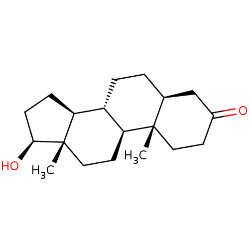
 DHT Dihydrotestosterone [3H]DHT (5alpha,17beta)-17-hydroxyandrostan-3-one BDBM50366473 (1S,2S,7S,10R,11S,14S,15S)-14-hydroxy-2,15-dimethyltetracyclo[8.7.0.0^{2,7}.0^{11,15}]heptadecan-5-one BDBM18161 CHEMBL27769
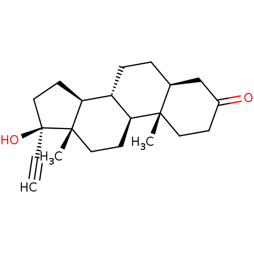 BDBM50423536 CHEMBL260202 17-Ethinyl-Dihydrotestosterone
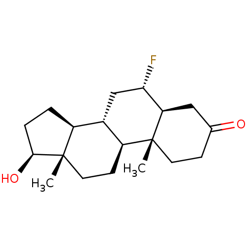 BDBM50423549 CHEMBL408707 6Alpha-Fluoro-Dihydrotestosterone
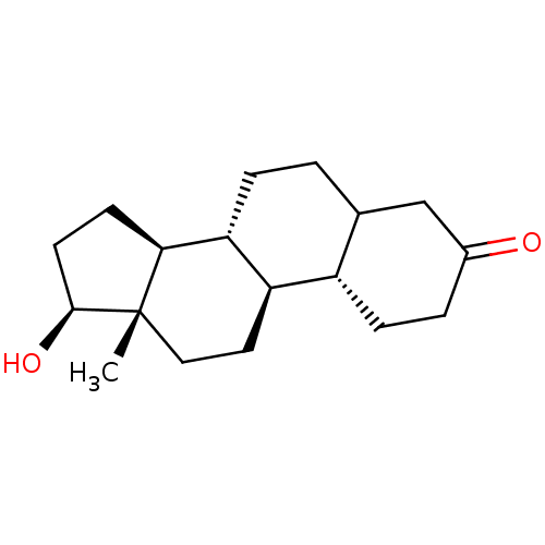 CHEMBL260641 19-Nor-Dihydrotestosterone BDBM50423543
 [3H]DHT BDBM25429
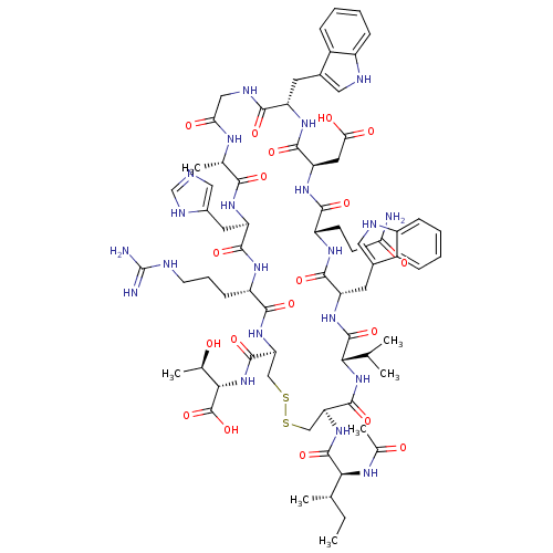 CHEMBL437340 Ac-I[CV(Dht)QDWGAHRC]T BDBM50159058
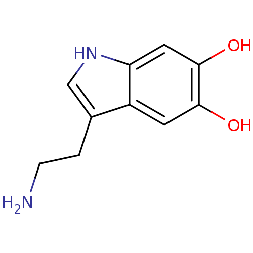 2-(5,6-Dihydroxy-1H-indol-3-yl)-ethyl-ammonium(5,6-DHT) CHEMBL303916 BDBM50012672 2-(5,6-Dihydroxy-1H-indol-3-yl)-ethyl-ammonium 3-(2-Amino-ethyl)-1H-indole-5,6-diol
- Schöttner, M; Spiteller, G; Gansser, D Lignans interfering with 5 alpha-dihydrotestosterone binding to human sex hormone-binding globulin. J Nat Prod 61: 119-21 (1998)
- Peřina, M; Kiss, MA; Mótyán, G; Szczyrbová, E; Eliáš, M; Študent, V; Kurfürstová, D; Kovalová, M; Mada, L; Bouchal, J; Frank, É; Jorda, R A-ring-fused pyrazoles of dihydrotestosterone targeting prostate cancer cells via the downregulation of the androgen receptor. Eur J Med Chem 249: (2023)
- Bellavance, E; Luu-The, V; Poirier, D Potent and selective steroidal inhibitors of 17beta-hydroxysteroid dehydrogenase type 7, an enzyme that catalyzes the reduction of the key hormones estrone and dihydrotestosterone. J Med Chem 52: 7488-502 (2009)
- ChEMBL_199192 (CHEMBL807120) In vitro inhibition of DHT (dihydrotestosterone) on proliferation of androgen-sensitive cancer Schionogi (SC-3) cells
- ChEBML_36274 Displacement of DHT from human androgen receptor
- ChEMBL_422532 (CHEMBL911029) Displacement of [3H]DHT from human SHBG
- BIOLOGICAL ACTIVITY Reagents and instruments: radiolabeled dihydrotestosterone (DHT-d3) and unlabelled dihydrotestosterone (DHT) purchased from Sigma-Aldrich (St. Louis, Mo.), scintillation solution purchased from Perkin Elmer Life Sciences (Boston, Mass.), hydroxyapatite (HAP) suspension purchased from Bio-Rad Laboratories (Hercules, Calif.), buffer (containing 10 mM Tris, 1.5 mM disodium EDTA, 0.25 M sucrose, 10 mM sodium molybdate and 1 mM PMSF, pH value adjusted to 7.4), and hydroxyapatite (HAP) solution (containing 50 mM Tris and 1 mM KH2PO4, pH value adjusted to 7.4).
- ChEMBL_36270 (CHEMBL648837) Displacement of [3H]DHT from human Androgen receptor
- ChEMBL_36271 (CHEMBL648838) Displacement of [3H]DHT from human Androgen receptor
- ChEMBL_547404 (CHEMBL1025921) Displacement of [3H]DHT from human serum SHBG
- ChEMBL_466706 (CHEMBL922711) Displacement of [3H]5alpha dihydrotestosterone from human sex hormone binding globulin
- ChEMBL_457372 (CHEMBL940963) Displacement of [3H]dihydrotestosterone from human androgen receptor expressed in Sf9 cells
- ChEMBL_447720 (CHEMBL896728) Displacement of [3H]DHT from human androgen receptor in MDA453 cells
- ChEMBL_457595 (CHEMBL923796) Displacement of [3H]DHT from androgen receptor expressed in Sf9 cells
- ChEMBL_464720 (CHEMBL933063) Displacement of [3H]DHT from human androgen receptor in MDA453 cells
- ChEMBL_1539997 (CHEMBL3737755) Inhibition of rat liver type 1 5alpha-reductase assessed as transformation of testosterone to dihydrotestosterone
- ChEMBL_1539998 (CHEMBL3737756) Inhibition of human prostate type 2 5alpha-reductase assessed as transformation of testosterone to dihydrotestosterone
- ChEMBL_36273 (CHEMBL648840) Binding affinity towards human androgen receptor (hAR), using dihydrotestosterone as radioligand for competitive binding assay
- ChEMBL_1735708 (CHEMBL4151244) Antagonist activity at human AR assessed as reduction in DHT-induced transactivation
- ChEMBL_303028 (CHEMBL830415) Inhibition of [3H]DHT binding to androgen receptor of MDA-453 cells
- ChEMBL_303055 (CHEMBL828881) Inhibition of [3H]-DHT binding to T877A androgen receptor of LNCaP cells
- ChEMBL_303250 (CHEMBL826376) Displacement of [3H]DHT from androgen receptor of human MDA-453 cells
- ChEMBL_449233 (CHEMBL899495) Displacement of [3H]DHT from human recombinant AR expressed in Sf9 cells
- ChEMBL_643540 (CHEMBL1212404) Displacement of [3H]DHT from AR in human MDA-MB-453 cells
- ChEMBL_1441437 (CHEMBL3373392) Inhibition of rat liver 5alpha-reductase type 1 assessed as conversion of [3H]testosterone to dihydrotestosterone
- ChEMBL_1441438 (CHEMBL3373987) Inhibition of human prostate 5alpha-reductase type 2 assessed as conversion of [3H]testosterone to dihydrotestosterone
- ChEMBL_699332 (CHEMBL1647250) Inhibition of human prostate 5alpha-reductase type 2 assessed as formation dihydrotestosterone from [4-14C] testosterone
- ChEMBL_1627004 (CHEMBL3869525) Displacement of [3H]DHT from androgen receptor in human MDA-MB-453 cells
- ChEMBL_1707211 (CHEMBL4058444) Displacement of [3H]-DHT from recombinant human androgen receptor expressed in CHO cells
- ChEMBL_213114 (CHEMBL873905) In vitro inhibition of DHT production in Hs 68 (human genital fibroblast) cells.
- ChEMBL_305616 (CHEMBL829479) Inhibitory concentration against [3H]-DHT binding to human sex hormone-binding globulin (SHBG)
- ChEMBL_36276 (CHEMBL648375) Displacement of DHT from human androgen receptor expressed in baculovirus SF-12 cells
- ChEMBL_566235 (CHEMBL964136) Displacement of [3H]DHT from human cloned androgen receptor expressed in Sf9 cells
- ChEMBL_971947 (CHEMBL2405557) Antagonist activity at human pSG5-AR assessed as inhibition of dihydrotestosterone-induced effect by reporter gene assay
- ChEMBL_2227411 (CHEMBL5140924) Displacement of [3H]DHT from Androgen receptor expressed in human MDA-MB-453 cells
- ChEMBL_36275 (CHEMBL648842) Inhibition of DHT binding to human androgen receptor expressed in baculovirus SF-12 cells
- ChEMBL_479690 (CHEMBL933554) Displacement of [3H]DHT from human Androgen receptor in human MDA-MB-453 cells
- ChEMBL_637915 (CHEMBL1169306) Displacement of [3H]DHT from human androgen receptor after 16 hrs by scintillation counting
- ChEMBL_791030 (CHEMBL1925691) Displacement of [3H]DHT from human androgen receptor expressed in Escherichia coli BL21 cells
- ChEMBL_501560 (CHEMBL987689) Displacement of [3H]DHT from human cloned androgen receptor expressed in insect Sf9 cell system
- ChEMBL_1448081 (CHEMBL3371986) Displacement of [3H]DHT from androgen receptor in human MDA-MB-435 cells after 30 mins
- ChEMBL_634399 (CHEMBL1118176) Displacement of [3H]DHT from human AR expressed in baculovirus Sf9 cells system by scintillation counting
- ChEMBL_1653051 (CHEMBL4002306) Inhibition of human prostate 5-alpha reductase 2 assessed as decrease in conversion of testosterone to dihydrotestosterone by chromatographic method
- ChEMBL_475674 (CHEMBL937560) Inhibition of wild type androgen receptor expressed in CV1 cells assessed as dihydrotestosterone-stimulated transactivation by CAT reporter gene assay
- ChEBML_36272 Binding affinity to the human androgen receptor (hAR), using [3H]DHT as radioligand in a competitive binding assay
- ChEMBL_204724 (CHEMBL807638) Ability to inhibit Steroid 5-alpha-reductase in rat using Isotope method [3H]T to [3H]-DHT]
- ChEMBL_432234 (CHEMBL914606) Antagonist activity at androgen receptor expressed in HeLa cells assessed as inhibition of dihydrotestosterone-induced transcriptional activity by reporter gene assay
- Androgen Receptor Binding Assay The competitive radio-ligand binding analysis was performed on human AR extracts from transfected Sf9 cells in the presence or absence of differing concentrations of test compound and a fixed concentration of 3H-dihydrotestosterone (3H-DHT) as tracer. AR bound 3H-DHT levels are determined in the presence of compounds and compared to levels of receptor specific binding when no competitor is present. Compound binding affinity to the human AR is expressed as the concentration of compound at which 50% of the maximum specific binding is inhibited (IC50).
- ChEMBL_36272 (CHEMBL648839) Binding affinity to the human androgen receptor (hAR), using [3H]DHT as radioligand in a competitive binding assay
- ChEMBL_449393 (CHEMBL899661) Antagonist activity at human androgen receptor expressed in HeLa cells assessed as inhibition of dihydrotestosterone induced transcriptional activity by reporter gene assay
- ChEMBL_457596 (CHEMBL923797) Antagonist activity at androgen receptor expressed in MDA-MB-453 cells assessed as inhibition of dihydrotestosterone-induced response in reporter gene assay
- ChEBML_205039 Inhibitory activity against Steroid 5-alpha-reductase type 2 as [3H]T to [3H]-DHT conversion human prostate nuclear membrane
- ChEMBL_1519754 (CHEMBL3624233) Displacement of [3H]-DHT from androgen receptor in human MDA-MB-453 cells after 90 mins by TopCount analysis
- ChEMBL_2192222 (CHEMBL5104582) Displacement of [3H]-DHT from recombinant androgen receptor (unknown origin) incubated for 3 hrs by liquid scintillation counter method
- ChEMBL_36106 (CHEMBL648071) Antagonistic activity against human androgen receptor (hAR) in CV-1 cells was determined as a function of maximal inhibition of dihydrotestosterone using cotransfection assay
- ChEMBL_805650 (CHEMBL1960484) Antagonist activity at androgen receptor expressed in Cos7 cells assessed as inhibition of dihydrotestosterone-induced luciferase activity after 24 hrs by reporter gene assay
- ChEMBL_501561 (CHEMBL987690) Antagonist activity at human androgen receptor expressed in human MDA-MB-231 cells assessed as inhibition of DHT-induced response
- ChEMBL_1449353 (CHEMBL3372081) Inhibition of Sprague-Dawley rat liver steroid 5-alpha-reductase assessed as inhibition of testosterone conversion to dihydrotestosterone incubated for 30 mins by HPLC method
- ChEMBL_474428 (CHEMBL934261) Antagonist activity at human AR ligand binding domain expressed in african green monkey COS7 cells in presence of 5-alpha-dihydrotestosterone by Gal4 hybrid assay
- ChEMBL_1511238 (CHEMBL3607996) Inhibition of androgen receptor in human LNCAP cells assessed as inhibition of DHT-induced KLK2 gene expression by Array Plate assay
- ChEMBL_1511239 (CHEMBL3607997) Inhibition of androgen receptor in human LNCAP cells assessed as inhibition of DHT-induced KLK3 gene expression by Array Plate assay
- ChEMBL_1511240 (CHEMBL3607998) Inhibition of androgen receptor in human LNCAP cells assessed as inhibition of DHT-induced SPDEF gene expression by Array Plate assay
- ChEMBL_772899 (CHEMBL1838302) Displacement of [3H]DHT from human recombinant androgen receptor-LBD expressed in Escherichia coli after 18 hrs by liquid scintillation counting
- ChEMBL_945929 (CHEMBL2341003) Displacement of [3H] ]5alpha-DHT from mouse androgen receptor expressed in monkey COS7 cells after 2 hrs by scintillation counting analysis
- ChEMBL_1735695 (CHEMBL4151231) Antagonist activity at human CMX-AR expressed in HEK293 cells assessed as reduction in dihydrotestosterone-induced transactivation activity after 24 hrs by luciferase reporter gene assay
- ChEMBL_1742330 (CHEMBL4158080) Antagonist activity at AR in mouse SC3 cells assessed as inhibition of DHT-induced cell proliferation after 3 days by CCK8 assay
- ChEMBL_1803399 (CHEMBL4275691) Displacement of Fluormone-DHT from rat GST-tagged rat AR LBD expressed in insect cells after 4 hrs by fluorescence polarization assay
- ChEMBL_1933747 (CHEMBL4479399) Inhibition of DHT-induced androgen receptor transactivation in human LNCaP cells after 24 hrs by luciferase reporter gene assay relative to control
- ChEMBL_752066 (CHEMBL1786230) Antagonist activity at wild type androgen receptor expressed in african green monkey CV1 cells assessed as inhibition of dihydrotestosterone-induced transcriptional activity by luciferase reporter gene assay
- ChEMBL_1621743 (CHEMBL3864026) Displacement of fluormone-DHT green from His/GST-tagged rat AR ligand binding domain after 4 to 8 hrs by fluorescence polarization assay
- ChEMBL_2207001 (CHEMBL5119709) Antagonist activity at DHT-induced Androgen receptor transcriptional activity in human HEK293 cells measured after 24 hrs by Steady-Glo reagent based assay
- ChEMBL_580463 (CHEMBL1052490) Antagonist activity at androgen receptor in human MDA-kb2 cells assessed as inhibition of DHT-induced luciferase activity by luciferase reporter gene assay
- ChEMBL_634400 (CHEMBL1118177) Antagonist activity at androgen receptor in human MDA-MB-453 cells coexpressing MMTV-ARE reporter gene assessed as inhibition of DHT-induced response
- ChEMBL_1803354 (CHEMBL4275646) Antagonist activity at AR in human LNCAP cells assessed as reduction in DHT-induced transcriptional activation after 24 hrs by luciferase reporter gene assay
- ChEMBL_1519755 (CHEMBL3624234) Antagonist activity at androgen receptor in human MDA-MB-453 cells assessed as inhibition of DHT-induced PSA expression by alkaline phosphatase reporter gene assay
- ChEMBL_2450772 Inhibition of DHT induced androgen receptor transcription in human LNCaP cells cotransfected with PSA-luc and renilla plasmid incubated for 24 hrs by Dual luciferase assay
- ChEMBL_796526 (CHEMBL1937841) Antagonist activity at human androgen receptor expressed in COS7 cells assessed as inhibition of DHT-induced luciferase activity after 24 hrs by reporter gene assay
- ChEBML_204897 Inhibitory activity against Steroid 5-alpha-reductase type 1 enzyme based on the conversion of [3H]T to [3H]-DHT in nuclear membrane preparations from human scalp
- ChEMBL_1803361 (CHEMBL4275653) Antagonist activity at AR in human LNCAP cells assessed as reduction in DHT-induced cell growth treated for every 2 days for 4 days by hemocytometry
- ChEMBL_2014930 (CHEMBL4668508) Antagonist activity at androgen receptor (unknown origin) expressed in human HeLa cells harboring AR3A-PSA-(ARE)4-Luc13 in presence of DHT by BrightGlo luciferase assay
- ChEMBL_2027527 (CHEMBL4681685) Inhibition of wild-type androgen receptor in human LAPC4 cells assessed as inhibition of DHT-induced luciferase activity measured after 24 hrs by luciferase reporter assay
- ChEMBL_624727 (CHEMBL1113403) Antagonist activity at AR in human MDA-kb2 cells co-transfected with MMTV-luc assessed as decrease in DHT-induced luciferase activity by reporter gene assay
- ChEMBL_993099 (CHEMBL2446674) Antagonist activity at androgen receptor in mouse SC3 cells assessed as inhibition of DHT-induced cell growth after 3 days by CCK-8/WST-8 assay
- ChEMBL_205061 (CHEMBL810255) Inhibitory activity against Steroid 5-alpha-reductase type I enzyme based on the conversion of [3H]-T to [3H]DHT in nuclear membrane preparations from human scalp
- ChEMBL_841247 (CHEMBL2089650) Antagonist activity at wild type AR LBD assessed as inhibition of DHT-induced fluorescent labeled D11-FxxLF recruitment after 2 to 4 hrs by TR-FRET assay
- ChEMBL_1520318 (CHEMBL3626368) Antagonist activity at wild type AR expressed in human SC cells assessed as inhibition of 1 nM DHT-induced cell proliferation after 3 days by WST-8 assay
- ChEMBL_1621736 (CHEMBL3864019) Antagonist activity at wild type AR in human PC3 cells assessed as suppression of DHT-induced receptor transcriptional activation after 24 hrs by PSA-luciferase reporter gene assay
- ChEMBL_2160360 (CHEMBL5045110) Binding affinity to human AR LBD (663 to 920 residues) expressed in Escherichia coli BL21 (DE3) assessed as dissociation constant in presence of DHT by biolayer interferometry assay
- ChEMBL_993098 (CHEMBL2446673) Displacement of [3H]-DHT from human GST-tagged androgen receptor LBD (627 to 919) expressed in Escherichia coli HB-101 after 15 hrs by liquid scintillation counting analysis
- SPA Binding Assay (IC50) and Coactivator Peptide Competitive FP Assay (IC50/EC50) A radiolabeled ligand competition scintillation proximity assay (SPA) for the androgen receptor (AR) using Ni-coated 384-well FlashPlates. It measures [3H]-DHT displacement from AR-LBD protein. AR functionality was determined by fluorescence polarization (excitation @485 nm, emission @ 530 nm) assay, which measures fluorescently labeled SRC2-3 peptide displacement from DHT-bound AR-LBD.
- ChEMBL_1520320 (CHEMBL3626370) Displacement of [3H]-DHT from human GST fused AR-LBD (627 to 919 amino acids) transfected in Escherichia coli HB 101 after 15 hrs by liquid scintillation counting assay
- ChEMBL_2031518 (CHEMBL4685676) Antagonist activity at androgen receptor in human LNCaP cells transfected with ARR2 PB-eGFP assessed as inhibition of DHT-induced transcriptional activity measured after 3 days by fluorescence assay
- ChEMBL_2325367 Antagonist activity at wild type androgen receptor expressed in HEK293 cells assessed as reduction in DHT-induced transcriptional activation of androgen receptor incubated for 24 hrs by Steady-Glo assay
- ChEMBL_2325369 Antagonist activity at androgen receptor F876L mutant expressed in HEK293 cells assessed as reduction in DHT-induced transcriptional activation of androgen receptor incubated for 24 hrs by Steady-Glo assay
- ChEMBL_2325371 Antagonist activity at androgen receptor W741L mutant expressed in HEK293 cells assessed as reduction in DHT-induced transcriptional activation of androgen receptor incubated for 24 hrs by Steady-Glo assay
- ChEMBL_2325373 Antagonist activity at androgen receptor T877A mutant expressed in HEK293 cells assessed as reduction in DHT-induced transcriptional activation of androgen receptor incubated for 24 hrs by Steady-Glo assay
- ChEMBL_2364315 Inhibition of androgen receptor in human VCaP cells expressing GRE-2-TATA-Luc co-expressing firefly luciferase incubated for 24 hrs in presence of DHT by luciferase reporter gene assay
- ChEMBL_1658408 (CHEMBL4008020) Antagonist activity at GAL4-fused human AR LBD (667 to 919 residues) expressed in CHO-K1 cells assessed as inhibition of dihydrotestosterone-induced transactivation activity after 5 to 6 hrs by luciferase reporter gene assay
- SPA Binding Assay (IC50) A radiolabeled ligand competition scintillation proximity assay (SPA) for the androgen receptor (AR) using Ni-coated 384-well FlashPlates. It measures [3H]-DHT displacement from AR-LBD protein.
- ChEMBL_513497 (CHEMBL975403) Antagonist activity at human androgen receptor expressed in mouse NIH3T3 cells assessed as inhibition of DHT-induced transcriptional activation after 24 hrs by androgen response element-mediated luciferase reporter gene assay
- ChEMBL_1678006 (CHEMBL4028149) Antagonist activity at human GAL4-DBD fused AR ligand binding domain transfected in human Huh7 cells co-expressing GAL4-RE-Luc assessed as reduction in dihydrotestosterone-induced luciferase activity after 16 hrs by luciferase reporter gene assay
- ChEMBL_2034916 (CHEMBL4689074) Antagonist activity at androgen receptor (unknown origin) expressed in HEK293 cells using DHT as substrate preincubated for 30 mins followed by substrate addition measured after 24 hrs by Steady-Glo luciferase assay
- Coactivator Peptide Competitive FP Assay (IC50/EC50) AR functionality was determined by fluorescence polarization (excitation @485 nm, emission @ 530 nm) assay, which measures fluorescently labeled SRC2-3 peptide displacement from DHT-bound AR-LBD.
- ChEMBL_941808 (CHEMBL2329799) Agonist activity at androgen receptor (unknown origin) expressed in HeLa cells co-expressing PSA-(ARE)4-Luc13 assessed as induction of DHT-induced luciferase activity after 20 mins by luciferase reporter gene assay
- ChEMBL_815556 (CHEMBL2025085) Antagonist activity at androgen receptor expressed in HeLa-AR3A-PSA-(ARE)4-Luc13 cells assessed as rightward shift of DHT-induced response incubated for 20 hrs at 37 degC by luciferase reporter gene assay
- 5α-Reductase Activity Assay The reaction mixture contained a final volume of 1 mL: 1 mM dithiothreitol, sodium phosphate buffer 40 mM, at pH 6.5 for human prostate, 2 mM NADPH, 2 nM [1,2,6,7-3H]T [Cabeza et al., Steroids 60:630-5]. The reaction in duplicate was started when it was added to the enzymatic fraction (500 μg protein in a volume of 80 μL) incubated at 37°C for 60 min [Hirosumi et al., J. Steroid Biochem. Mol. Biol., 52:357-63] and stopped by mixing with 1 mL of dichloromethane; this was considered as the end point. Incubation without tissue was used as a control. The mixture (incubation medium/dichloromethane) was agitated on a vortex for 1 min and the dichloromethane phase was separated and placed in another tube. This procedure was repeated four more times. The dichloromethane extract was evaporated to dryness under a nitrogen stream and suspended in 50 mL of methanol that was spotted on high-performance thin layer chromatography (HPTLC) Keiselgel 60 F254 plates. T and DHT were used as carriers, and were applied in different lanes on both lateral sides of the plates (T, T + DHT, and DHT). The plates were developed in chloroform-acetone, 9:1, and were air-dried; the chromatography was repeated twice more. The steroid carriers were detected using phosphomolybdic acid reagent (DHT) and with an ultraviolet (UV) lamp (254 nm) (T). After the plates were segmented into pieces of 1 cm each, these were cut off and the strips soaked in 5 mL of Ultima Gold (Packard). The radioactivity was determined in a scintillation counter (Packard Tri-Carb 2100 TR). The radioactivity content in the segment corresponding to T and DHT carriers was identified. Radioactivity with identical chromatographic behavior to the DHT standard was considered as the DHT transformation. Control incubations, chromatography separations, and identifications were carried out in the same manner as described above, except that the tubes did not contain tissue.
- Receptor Binding and Transactivation Assay Receptor Binding Assay (Ki)-Binding determined through direct displacement of ligand with [3H]-DHT in the MDA-453 cell line. Transactivation Assay (EC50, IC50) - Functional agonist/antagonist activities determined through a stably transfected mouse myoblast C2C12 cell line utilizing a luciferase reporter.
- 5α-R1 Activity Assay The activity of the 5α-R type 1 isozyme was determined by following the conversion of T to DHT at pH 7.5. 2 nM of [1,2,6,7 3H] T, and different concentrations of unlabeled T (5 × 10^-7-6.3 × 10^-6 M) or DHT (5 × 10^-7-2.7 × 10^-5M) and 2 mM NADPH11 was prepared. The reaction in duplicate was started when 5α-R type 1 isozyme fraction (60 μg protein in a volume of 7.5 μL) was added to the mixtureand incubated at 37°C for 60 min.
- 5α-Reductase Activity Assay The reaction mixtures contained a final volume of 1 mL, 1 mM DTT, sodium phosphate buffer 40 mM, at pH 6.5, 2 mM, NADPH, 2 nM [1,2,6,7-3H]T or [1,2,3,7-3H(N)] 4-dione and the prostatic enzyme fractions. The amount of prostatic enzyme fractions was determined to adjust the rate of conversion of T to DHT or 4-dione to 5α-dione to around 28%. T, DHT, unlabeled 4-dione or 5α-dione concentrationswere adjusted to 25-250 nM in the incubating medium, by adding one of the following reagents cold T, DHT, unlabeled 4-dione or 5α-dione. The reactions in duplicate were started when added to the enzymatic fraction (400 μg protein in a volume of 80 μL) incubated at 37°C for 60 min13 and stopped by mixing with 1 mL ofdichloromethane. Incubation without tissue was used as a control. All reactions were carried out in two different times by duplicate.
- Antagonist Activity for AR Antagonist activity for AR was evaluated according to the following method. COS-7 cells (ATCC) were transfected with pMMTV-luc vector (reporter plasmid having, as an androgen response element, murine mouse mammary virus long terminal repeat) and pEX-hAR vector (human androgen receptor expression vector: which expresses human AR gene under control of CMV promoter) by using Nucleofector (registered trademark) Kit R (Lonza) as a transfection reagent and Amaxa (Lonza). The COS-7 cells obtained after transfection were seeded in a clear bottom 96 well microplate (BD) at 1.5×10⁴/well with phenol red free RPMI1640 containing 10% charcoal-treated fetal bovine serum (hereinbelow, DCC-FBS) (hereinbelow, the medium is referred to as an evaluation medium), and then cultured overnight. The culture was added with the evaluation medium containing dihydrotestosterone (DHT) (final concentration of DHT: 1 nmol/L) or the evaluation medium containing the compound of Examples or the compound of Comparative Examples (final concentration of the compound of Examples or the compound of Comparative Examples: 5, 14, 41, 123, 370, 1111, 3333, or 10000 nmol/L), followed by culture for 24 hours. Then, the transcription activity value was measured. The transcription activity was measured by using Bright-Glo Luciferase Assay System (Promega). From the measured transcription activity, 50% transcription activity inhibition concentration (IC50 value) was calculated by logistic regression when the transcription activity value obtained by using 1 nmol/L DHT was 100% and the transcription activity value obtained by using the evaluation medium only was 0%.
- Mammalian Two-Hybrid Assay We performed a mammalian one-hybrid assay in HeLa cells, where we transfected a construct coding for a Gal4DBD-BagN128 fusion or just Gal4DBD alone togetherwith a Gal4 binding site-luciferase reporter gene and an AR expression vector. The transfected cells were then treated with or without DHT, and the reporter gene activity was measured.
- In Vitro Inhibition Assay This work was performed at the MDS Pharma Services, Pharmacology Laboratories, Taiwan. The assay was an in vitro evaluation of the ability of an extract or a pure compound to inhibit the steroid 5alpha -reductase enzyme from metabolizing testosterone into dihydrotestosterone. This is an enzyme-immunoassay (EIA) for quantitative determination of testosterone in human serum or plasma. The significance of this type of inhibition is that it can lead to eradication of benign prostatic hyperplasia (BPH). Two distinct isozymes are found in mice, rats, monkeys and humans: type 1 and II. Each of these isozymes is differentially expressed in tissues and developmental stages. In human, type 1 steroid 5alpha -reductase is predominant in the sebaceous glands of most regions of skin, including scalp and liver and is responsible for approximately one third of circulating DHT. Inhibitors of steroid 5alpha -reductase may be of benefit in the treatment of androgenetic alopecia.
- Luciferase Reporter Assay Activities of the test compounds to inhibit androgen receptor mediated transcription induced by DHT were evaluated using stable transfected AR/CHO#3 cells plated onto 96-well luminoplates. After cells were lysed, cell extracts were prepared for luciferase assay. The luciferase activity was measured with a ML3000 luminometer. AR antagonist activities were calculated. The concentration of compounds showing 50% AR antagonist activity, the IC50 values, were obtained through nonlinear analysis using the statistical analysis system (SAS).
- Receptor Binding Assay (Ki) and Transactivation Assay (EC50) Binding assays were conducted by incubating test compound at various concentrations with [3H]DHT with human cancer epithelial breast cell lines MDAMB-45. IC50 value was obtained from dose-response displacement curves. The inhibition constant (Ki) was calculated from IC50 value by the Cheng and Prusoff equation. EC50 value was obtained from functional transactivation assay, which was conducted to assess the potency and efficacy of agonist/antagonist in stably transfected mouse myoblast C2C12 cell line utilizing a luciferase reporter.
- Binding Activity of Compound to Androgen Receptor AR The binding activity of the compound to AR was determined using the LanthaScreen TR-FRET Androgen Receptor Coactivator Assay kit (brand: Thermo, Cat. No: A15878). The AR receptor agonist dihydrotestosterone (DHT) (brand: Sigma, Cat. No: D-073) was diluted 10-fold with DMSO in a 96-well V-bottom plate. The highest concentration was 100 μM, 8 concentrations in total. The test compound was diluted 10-fold with DMSO. The highest concentration was 3000 μM, 8 concentrations in total. The compound was further diluted 50-fold with the detection buffer Nuclear Receptor Buffer A (containing 5 mM DTT) provided in the kit, 10 μL of the diluted compound was transferred to a 96-well half-area microplate, 5 μL of AR-LBD protein (4× concentration) was added to the test compound, then a mixture of 5 μL of fluorescein-coactivator peptide and Tb-labeled anti-GST antibody (4× concentration) was added, and the experiment was performed in duplicate. The plate was incubated at room temperature for 2 h, the fluorescence values (Excitation 337, Emission 520/495 nm) were detected using the PHERAstar instrument, and the 520:495 ratio was calculated. With the log value of the final concentration of the compound as the X axis and the 520/495 ratio as the Y axis, data was input into the processing software Graphpad Prism 9, and the EC50 value was calculated by four-parameter fitting.
- Inhibition Assay Inhibitors were initially screened for an ability to block the NADP+ dependent oxidation of the artificial substrate S-tetralol catalyzed by AKR1C3. S-tetralol was used since it is also a substrate of the highly related AKR1C1 and AKR1C2 enzymes. Inhibition of AKR1C1 and AKR1C2 is undesirable in the context of prostate cancer since they are involved in the elimination of DHT (see FIG. 1B; see also Rizner et al, 2003; Steckelbroeck et al. 2004). All assays were performed at Km so that IC50 values across enzyme forms were comparable.An exemplary screening strategy was against homogeneous recombinant AKR1C3 and its highly related enzyme AKR1C2 to generate full dose-response curves and IC50 values.
- Fluorescence Polarization Assay For the fluorescence polarization experiments, 5 nM fluorescently labeled Bag-1L (Bag-1L(1-20), fluorescein isothiocyanate (FITC)-MAQRGGARRPRGDRERLGSR; Bag-1L(61-80), FITC-RGAAAGARRPRMKKKTRRRS) or SRC-2 peptide (FITC-HDSKGQTKLLQLLTTKSDQM) was incubated with serially diluted AR-LBD (4 to 0.4 μM) in binding buffer (50 mM NaPO4, pH 6.5, 50 mM KCl, 1 mM DTT) and 10 μM DHT. The samples were then equilibrated for 45 min at ambient temperature. For the competition experiments, 400 to 0.4 nM Bag-1L core (GARRPR) or SRC-2 peptides (HDSKGQTKLLQLLTTKSDQM) were incubated with 4 μM AR-LBD and 5 nM fluorescent Bag-1 or SRC-2 peptide. Binding was measured using polarization (excitation λ = 485 nm, emission λ = 530 nm) on a Synergy H1 hybrid reader (Biotek).
- Gal4-luciferase reporter assay Briefly, the AR LBD is expressed as a fusion with the Gal4 transcription factor, which binds to the Gal4 reporter DNA. Upon activation with agonist hormone (DHT), the Gal-AR LBD binds to the Gal4 reporter gene, which in turn drives the transcription (and subsequently translation) of the reporter luciferase. The effect of antiandrogens (competitive antagonists) on this process is measured by quantifying the amount of luciferase activity after 24 hours in the presence of varying concentrations of antiandrogens. From these values, an IC50 for each antagonist is calculated. Experimentally, Hela cells were maintained in Dulbecco's modified Eagle's medium H-21 4.5 g/L glucose, containing 10% steroid depleted fetal bovine serum, 50 units/mL penicillin. For transfection, (1×10⁵) cells per well were plated and incubated overnight. A mixture of typically 200 ng of Gal4 responsive luciferase reporter plasmid, 10 ng of β-actin-β-galactosidase internal control, 10 ng of GAL-AR LBD or empty vector control, and 10-100 ng of β-catenin or empty vector control were mixed with 0.5 μL of transfection reagent from BioRad and incubated for 20 min and then plated in 24 well plate triplicates. Cells were induced with 1 nM DHT and 0.1 nM-30 uM test compounds after 3 hr and then incubated over night. Cells were collected, and pellets were lysed in 100 μL of 100 mM Tris-HCl (pH 7.5) containing 0.1% Triton X-100. Luciferase and β-galactosidase activities were measured using the Luciferase Assay System (Promega) and Galacto-Light Plus-galactosidase reporter gene assay system (Applied Biosystems), according to the manufacturer's instructions.
- Discontinuous Radiometric Assay Compounds may be evaluated as selective reversible inhibitors of AKR1C3 by screening them against homogeneous recombinant AKR1C1-AKR1C4 expressed in E. coli. In each case, a discontinuous radiometric assay may be used to monitor the inhibition of progesterone reduction (20-ketosteroid reduction) catalyzed by AKR1C1, the inhibition of Δ4-AD reduction (17-ketosteroid reduction) catalyzed by AKR1C3, and the inhibition of 5α-DHT reduction (3-ketosteroid reduction) catalyzed by AKR1C2 and AKR1C4 (by measuring the formation of 20α-hydroxyprogesterone, testosterone or 3α-androstanediol by radiochromatography). Secondary screens of the compounds of interest include: (a) a full-screen against all nine human recombinant AKR enzymes to ensure there are no-intended off-target effects (in this context AKR1B10 (retinal reductase; SEQ ID NO:5) has been shown to be potently inhibited by N-phenylanthranilates) (Endo et al., 2010, Biol. Pharm. Bull. 33:886-90); (b) a screen against COX-1 and COX-2 to reaffirm that compounds do not act as NSAIDs; and (c) an expanded screen against other nuclear receptors (especially other steroid hormone receptors).
- Human Androgen Receptor (hAR) Ligand Binding Domain (LBD) Affinity Assay Methods: hAR-LBD (633-919) was cloned into pGex4t.1. Large scale GST-tagged AR-LBD was prepared and purified using a GST column. Recombinant AR-LBD was combined with [3H]mibolerone (PerkinElmer, Waltham, Mass.) in buffer A (10 mM Tris, pH 7.4, 1.5 mM disodium EDTA, 0.25 M sucrose, 10 mM sodium molybdate, 1 mM PMSF) to determine the equilibrium dissociation constant (Kd) of [3H]mibolerone. Protein was incubated with increasing concentrations of [3H]mibolerone with and without a high concentration of unlabeled mibolerone at 4° C. for 18 h in order to determine total and non-specific binding. Non-specific binding was then subtracted from total binding to determine specific binding and non-linear regression for the ligand binding curve with one site saturation was used to determine the Kd of mibolerone.Increasing concentrations of SARDs or DHT (range: 10−12 to 10−4 M) were incubated with [3H]mibolerone and AR-LBD using the conditions described above. Following incubation, the ligand bound AR-LBD complex was isolated using BiogelHT hydroxyapatite, washed and counted in a scintillation counter after adding scintillation cocktail.
- mmTV-luciferase reporter assay An AR-response element is contained within the mmTV sequence and drives the expression of luciferase. The effect of antiandrogens (competitive antagonists) on this process is measured by quantifying the amount of luciferase activity after 24 hours in the presence of varying concentrations of antiandrogens. From these values, an IC50 for each antagonist is calculated. Experimentally, HeLa cells were maintained in Dulbecco's modified Eagle's medium H-21 4.5 g/L glucose, containing 10% steroid depleted fetal bovine serum, 50 units/mL penicillin. For transfection, (1×10⁵) cells per well were plated and incubated overnight. A mixture of typically 200 ng of mmTV responsive luciferase reporter plasmid, 10 ng of β-actin-β-galactosidase internal control, 10 ng of AR full length CMV expression vector or empty vector control, and 10-100 ng of β-catenin or empty vector control were mixed with 0.5 μL of transfection reagent from BioRad and incubated for 20 min and then plated in 24 well plate triplicates. Cells were induced with 1 nM DHT and 0.1 nM-30 uM compounds after 3 hrs and then incubated overnight. Cells were collected, and pellets were lysed in 100 μL of 100 mM Tris-HCl (pH 7.5) containing 0.1% Triton X-100. Luciferase and β-galactosidase activities were measured using the Luciferase Assay System (Promega) and Galacto-Light Plus-galactosidase reporter gene assay system (Applied Biosystems), according to the manufacturer's instructions.